12 Cranial Nerves
cranial nerves, trigeminal nerve, cranial nerves, maxillary nerve, trigeminal nerve branches, cranial nerves, 12 cranial nerves, cranial nerves, zygomatic nerve, olfactory nerve, infraorbital nerve, cranial nerves, cranial nerves, CRANIAL NERVES, branches of trigeminal nerve, pterygopalatine ganglion, eye anatomy, trigeminal nerve anatomy, superior alveolar nerve, cranial nerves,
The following are the result pages for the searched keyowrd: 12 Cranial Nerves

Cranial nerves anatomy
22/10/2009 01:51:53 م
In This Section you will find detailed different Photos and images about the anatomy of the Cranial Nerves including Their types , Fascial nerve anatomy , trigeminal nerve anatomy , vagus nerve anatom... More Details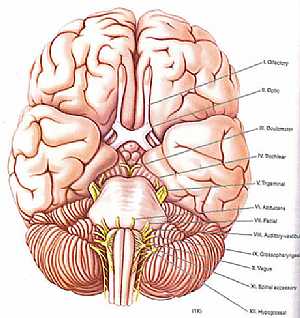
cranial nerves anatomy
14/10/2009 04:29:00 ص
a colored image for the cranial nerves exit showing: 1. olfactory nerve 2. optic nerve 3. occulomoto rnerve 4. trochlear nerve 5. trigeminal nerve 6. abducent nerve 7. fascial nerve 8. vestibulocochle... More Details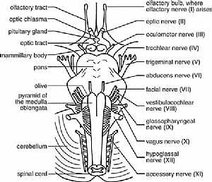
cranial nerves anatomy
14/10/2009 04:16:00 ص
this diagram shows the 12 cranial nerves and where they exactly exit from the brain showing: 1. all cranial nerves 2. olfactory bulb 3. optic chisma 4. pituitary gland 5. optic tract 6. mammillary bod... More Detailscranial nerves anatomy
14/10/2009 04:16:00 ص
this diagram shows the 12 cranial nerves and where they exactly exit from the brain showing: 1. all cranial nerves 2. olfactory bulb 3. optic chisma 4. pituitary gland 5. optic tract 6. mammillary bod... More Details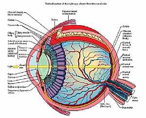
Eye anatomy
11/10/2009 04:00:00 م
this diagram details the different parts and structures of the human eye showing: 1. conjunctiva 2. ora serrata 3. cilliary body 4. aqueous 5. iris 6. ant. chamber 7. cornea 8. pupil 9. lens 10. post.... More DetailsCranial nerves anatomy
05/11/2009 04:03:00 ص
this image shows the all cranial nerves and displaying their effector organs showing: 1. Olfactory nerve I 2. Optic nerve II 3. Occulomotor nerve III 4. Trochlear nerve IV 5. Trigeminal nerve V 6. Ab... More Details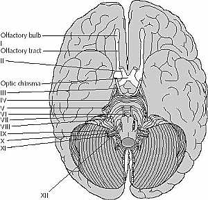
cranial nerves anatomy
14/10/2009 04:17:00 ص
this image is an inferior view of the brain showing the cranial nerves showing: 1. olfactory nerve 2. optic nerve 3. occulomoto rnerve 4. trochlear nerve 5. trigeminal nerve 6. abducent nerve 7. fasci... More Detailscranial nerves anatomy
14/10/2009 04:17:00 ص
this image is an inferior view of the brain showing the cranial nerves showing: 1. olfactory nerve 2. optic nerve 3. occulomoto rnerve 4. trochlear nerve 5. trigeminal nerve 6. abducent nerve 7. fasci... More Details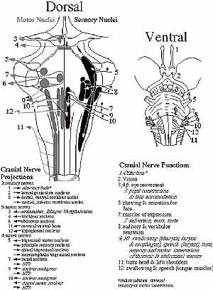
cranial nerves anatomy
14/10/2009 04:28:00 ص
this diagram shows location of the cranial nerves nuclei and their site of exit the right image is an anterior view and the left is a posterior view showing: 1. olfactory nerve 2. optic nerve 3. occul... More Details
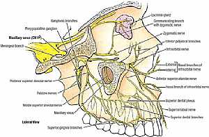
Trigeminal nerve anatomy
28/10/2009 01:55:00 ص
this images illustrates the different branches of the trigeminal nerve in the face in relation to each other [focusing on the maxillary division] showing: 1. maxillary nerve 2. meningeal branch 3. po... More Details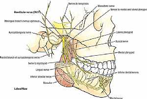
Trigeminal nerve anatomy
28/10/2009 02:07:00 ص
this images illustrates the different branches of the trigeminal nerve in the face in relation to each other [focusing on the mandibular division] showing: 1. mandibular nerve 2. meningeal branch 3. ... More Details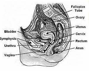
Vagina anatomy
13/11/2009 06:27:00 ص
this image shows the vagina of the female pelvis in relation to the surrounding organs this is a side view of a vertically sectioned pelvis showing: 1. fallopian tube 2. ovary 3. uterus 4. cervix 5. r... More Detailsorthopaedic joint assessment centr dr mcmahon
, , , , , , ,anatomi ligamen panggul wanita
, ,abdomen sans preparation normale
, , , , , ,world conferences on urine therapy
, , , , , , , , , ,© Copyright 2001-2022 eDoctorOnline.com