Base of the Skull
cranial nerves, jugular foramen, carotid canal, foramen lacerum, skull anatomy, base of skull, base of the skull, chiasmatic groove, hypoglossal canal, foramen magnum, tuberculum sellae, skull base, cranial nerves skull, cranial nerves skull, base of skull, base of the skull, Base of the Skull, base of the skull, cranial nerves in skull, mastoid foramen,
The following are the result pages for the searched keyowrd: Base of the Skull
Base of the skull. Upper surface
09/10/2009 03:29:00 م
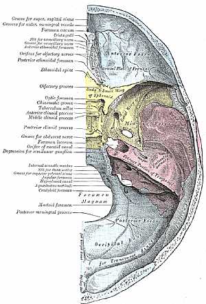
detailed image of the superior view for the base of the skull (ant. ,middle, and posterior cranial fossa with their structures and most important features) showing: 1. groove for superior sagital sinu... More Details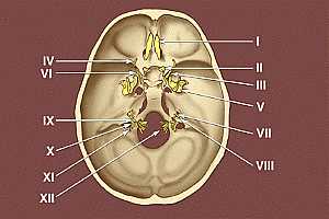
Base of the skull
09/10/2009 03:34:00 م
this image shows the exit of the twelve cranial nerves from the base of the skull * I. Olfactory nerve * II. Optic nerve * III. Oculomotor nerve * IV. Trochlear nerve * V. Trigeminal nerve * VI.... More Details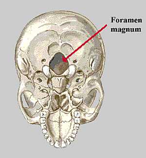
Base of the skull
10/10/2009 04:22:00 م
this image shows what is called foramen magnum the biggest opening in the base of the skull for the transmission of the spinal cord... More Details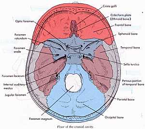
Base of the skull
10/10/2009 04:25:00 م
this a detailed image for the floor of the cranial cavity showing: 1. groove for superior sagital sinus 2. groove for ant. meningeal vessels 3. foramen caecum 4. crista gali 5. slit for nasociliary ne... More Details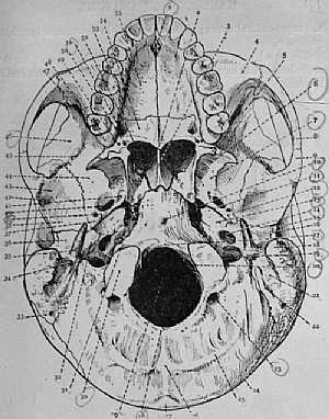
Base of the skull
24/10/2009 03:11:00 م
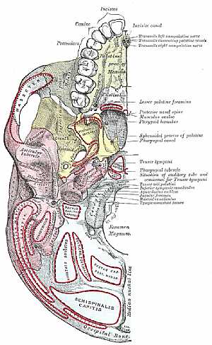
this is the base of the skull from below showing: 1. incisors 2. canines 3. premolars 4. molars 5. incisive canal 6. lesser palatine foramina 7. post. nasal spine 8. musculus uvulae 9. ptrygoid hamul... More DetailsBase of the skull
10/10/2009 04:25:00 م
this a detailed image for the floor of the cranial cavity showing: 1. groove for superior sagital sinus 2. groove for ant. meningeal vessels 3. foramen caecum 4. crista gali 5. slit for nasociliary ne... More Details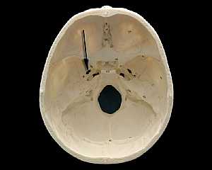
Skull Anatomy
19/10/2009 03:02:24 م
In This Section you will find detailed different Photos and images about the anatomy of the Skull bone including its surface , attachments related structures many more Items about the Skull anatomy... More DetailsBase of the skull
24/10/2009 03:11:00 م
this is the base of the skull from below showing: 1. incisors 2. canines 3. premolars 4. molars 5. incisive canal 6. lesser palatine foramina 7. post. nasal spine 8. musculus uvulae 9. ptrygoid hamul... More Details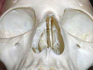
Skull Anatomy
10/10/2009 04:24:00 م
this is an anterior view of the skull showing two orbital cavities above and the nasal cavity below... More DetailsBase of the skull
10/10/2009 04:22:00 م
this image shows what is called foramen magnum the biggest opening in the base of the skull for the transmission of the spinal cord... More Details
Base of skull. Inferior surface
09/10/2009 03:53:00 م
this is a detailed inferior view for the base of the skull showing: 1. incisors 2. canines 3. premolars 4. molars 5. incisive canal 6. lesser palatine foramina 7. post. nasal spine 8. musculus uvulae ... More Details
Base of the skull
09/10/2009 03:34:00 م
this image shows the exit of the twelve cranial nerves from the base of the skull * I. Olfactory nerve * II. Optic nerve * III. Oculomotor nerve * IV. Trochlear nerve * V. Trigeminal nerve * VI.... More DetailsBase of skull. Inferior surface
09/10/2009 03:53:00 م
this is a detailed inferior view for the base of the skull showing: 1. incisors 2. canines 3. premolars 4. molars 5. incisive canal 6. lesser palatine foramina 7. post. nasal spine 8. musculus uvulae ... More Details
Hypothalamus and Thalaums anatmy
22/10/2009 01:50:32 م
In This Section you will find detailed different Photos and images about the anatomy of the Thalamus and Hypothalamus including its surface , parts , related structures , Blood supply and many more It... More Detailsorthopaedic joint assessment centr dr mcmahon
, , , , , , ,anatomi ligamen panggul wanita
, ,abdomen sans preparation normale
, , , , , ,world conferences on urine therapy
, , , , , , , , , ,© Copyright 2001-2022 eDoctorOnline.com