GLOSSOPHARYNGEAL nerve
cranial nerves, cranial nerves, ansa cervicalis, cranial nerves, hypoglossal nerve, 12 cranial nerves, vagus nerve, olfactory nerve, cranial nerves, taste pathway, cranial nerves, CRANIAL NERVES, base of skull, skull anatomy, cranial nerve anatomy, skull base, cranial nerves skull, optic nerve, image, base of the skull,
The following are the result pages for the searched keyowrd: GLOSSOPHARYNGEAL nerve
Glossopharybgeal nerve anatomy
30/10/2009 03:25:00 م
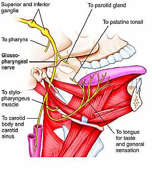
this image shows the glossopharyngeal nerve in the lateral aspect of the face displaying its course , branches and the related structures of the nerve showing: 1. superior ganglion 2. inferior gangli... More Details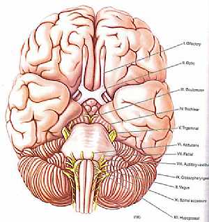
cranial nerves anatomy
14/10/2009 04:29:00 ص
a colored image for the cranial nerves exit showing: 1. olfactory nerve 2. optic nerve 3. occulomoto rnerve 4. trochlear nerve 5. trigeminal nerve 6. abducent nerve 7. fascial nerve 8. vestibulocochle... More Details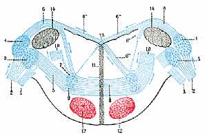
Vestibualr nerve anatomy
28/10/2009 03:44:00 م
this is 1. Vestibular nerve, divided at its entrance into the medulla oblongata. 2. Cochlear nerve. 3. Accessory nucleus of acoustic nerve. 4. Tuberculum acusticum. 5. Efferent fibers of accessory nu... More Details
Cranial nerves anatomy
22/10/2009 01:51:53 م
In This Section you will find detailed different Photos and images about the anatomy of the Cranial Nerves including Their types , Fascial nerve anatomy , trigeminal nerve anatomy , vagus nerve anatom... More Details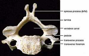
Vertebral Anatomy
19/10/2009 03:02:39 م
In This Section you will find detailed different Photos and images about the anatomy of the Vertebrae bones including their types , their surface , attachments related structures many more Items about... More Details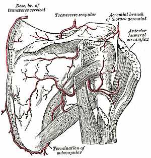
Blood supply of the upper limb
04/12/2009 06:47:00 ص
this image shows the arteries of the scapular region from posterior view (the arteries present on the scapula posteriorly) in relation to each other and to the surrounding bones and muscles showing: ... More Details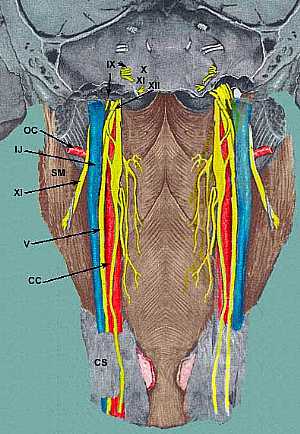
Cranial nerves anatomy
01/11/2009 02:27:00 م
this image shows the cranial nerves IX,X,XI,XII (glossopharyngeal , vagus , accessory and hypoglossal nerves) in relation to each other at the lateral aspect of the neck (pharynx) showing: 1. glossop... More Details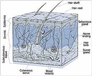
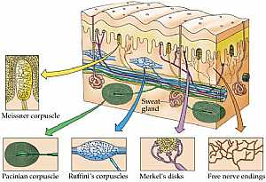
Skin sensation anatomy
03/11/2009 03:06:00 م
this image displays the different receptors present in the skin for different types of sensations showing: 1. pacinian corpusle "for pain and pressure sense" 2. ruffini's corpusles "s... More Details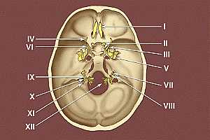
Base of the skull
09/10/2009 03:34:00 م
this image shows the exit of the twelve cranial nerves from the base of the skull * I. Olfactory nerve * II. Optic nerve * III. Oculomotor nerve * IV. Trochlear nerve * V. Trigeminal nerve * VI.... More Details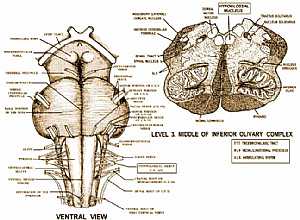
Hypoglossal nerve anatomy
03/11/2009 02:53:00 م
this image shows the hypoglossal nerve at its origin in relation to the surrounding structures showing: 1. hypoglossal nerve 2. cranial root of spinal accessory nerve 3. spinal root of spinal accesso... More Details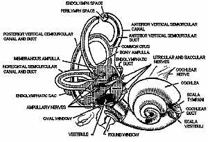
inner ear anatomy
12/10/2009 04:28:00 ص
this is a detailed image for the inner ear showing: 1. endolymph space 2. perilymph space 3. post. vertical semicircular canal and duct 4. membranous ampulla 5. horizontal semicircular canal and duct ... More Details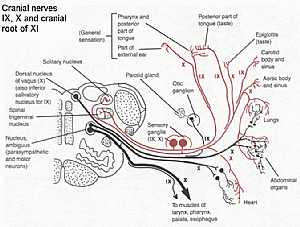
Cranial nerves IX,X,XI anatomy
28/10/2009 02:15:00 ص
this diagram shows the course of the cranial nerves( glossopharygeal IX,vagus X,and accessory XI ) from their origin to the their supplying organs showing: 1. solitary nucleus 2. spinal trigeminal nu... More Details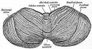
Cerebellum anatomy
24/10/2009 02:59:52 م
In This Section you will find detailed different Photos and images about the anatomy of the Cerebellums including their surfaces , parts , related structures , Functions , Blood supply of the Cerebell... More Details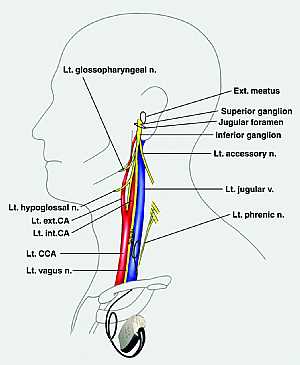
Glossopharybgeal nerve anatomy
29/10/2009 03:23:00 م
this is an image of the lateral side of the neck displaying the left glossopharyngeal nerve related to the surrounding structures showing: 1. left glossopharyngeal nerve 2. external auditory meatus 3... More DetailsGlossopharybgeal nerve anatomy
30/10/2009 03:25:00 م
this image shows the glossopharyngeal nerve in the lateral aspect of the face displaying its course , branches and the related structures of the nerve showing: 1. superior ganglion 2. inferior gangli... More Details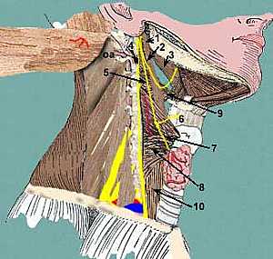
Glossopharybgeal nerve anatomy
28/10/2009 03:50:00 م
this image shows the glossopharyngeal nerve with the surrounding structures related to it in the area of the lateral aspect of the head and neck showing: 1. stylopharyngeus muscle 2. glossopharyngeal ... More DetailsCranial nerves anatomy
05/11/2009 04:03:00 ص
this image shows the all cranial nerves and displaying their effector organs showing: 1. Olfactory nerve I 2. Optic nerve II 3. Occulomotor nerve III 4. Trochlear nerve IV 5. Trigeminal nerve V 6. Ab... More DetailsHypoglossal nerve anatomy
03/11/2009 02:53:00 م
this image shows the hypoglossal nerve at its origin in relation to the surrounding structures showing: 1. hypoglossal nerve 2. cranial root of spinal accessory nerve 3. spinal root of spinal accesso... More Details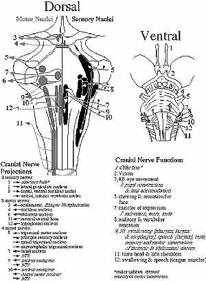
cranial nerves anatomy
14/10/2009 04:28:00 ص
this diagram shows location of the cranial nerves nuclei and their site of exit the right image is an anterior view and the left is a posterior view showing: 1. olfactory nerve 2. optic nerve 3. occul... More Details
Atlas of Human Anatomy
27/04/2006
In This Section you will find detailed different sections about all different parts of the human body including head and neck , chest , abdomen , upper limbs , pelvis and lower limbs and many more Sec... More Details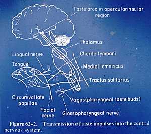
Taste sensation pathway
05/11/2009 03:59:00 ص
this image shows the pathway of the taste sensation to the cerebral cortex showing: 1. tongue 2. taste buds 3. lingual nerve 4. fascial nerve 5. glossopharyngeal nerve 6. vagus nerve 7. chorda tympan... More Details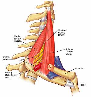
Neck Anatomy
19/10/2009 03:05:59 م
In This Section you will find detailed different Photos and images about the anatomy of the Neck including its surface , attachments , structures , Neck arteries , Neck Veins , trachea , Esophagus an... More DetailsTaste sensation pathway
05/11/2009 03:59:00 ص
this image shows the pathway of the taste sensation to the cerebral cortex showing: 1. tongue 2. taste buds 3. lingual nerve 4. fascial nerve 5. glossopharyngeal nerve 6. vagus nerve 7. chorda tympan... More Details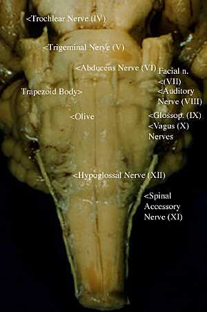
Nuclei of the cranial nerves
01/11/2009 02:45:00 م
this image shows a post view of the brain stem displaying the nuclei of the cranial nerves showing: 1. trochlear nerve 2. trigeminal nerve 3. abducent nerve 4. trapezoid body 5. olive 6. hypoglossal ... More DetailsNuclei of the cranial nerves
01/11/2009 02:45:00 م
this image shows a post view of the brain stem displaying the nuclei of the cranial nerves showing: 1. trochlear nerve 2. trigeminal nerve 3. abducent nerve 4. trapezoid body 5. olive 6. hypoglossal ... More Details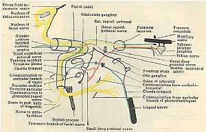
Fascial Nerve anatomy
01/11/2009 02:38:00 م
this diagram displays the course and branches of the fascial nerve showing: 1. fascial nerve nucleus 2. internal auditory meatus 3. small superfascial petrosal nerve 4. tympanic plexus 5. chorda tymp... More Detailscranial nerves anatomy
14/10/2009 04:29:00 ص
a colored image for the cranial nerves exit showing: 1. olfactory nerve 2. optic nerve 3. occulomoto rnerve 4. trochlear nerve 5. trigeminal nerve 6. abducent nerve 7. fascial nerve 8. vestibulocochle... More Details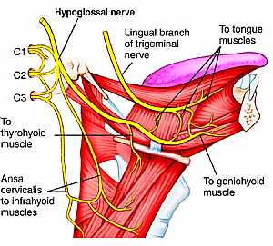
Hypoglossal nerve anatomy
03/11/2009 02:13:00 م
this image shows the cranial nerve XII "hypoglossal nerve" in the face region in the lateral aspect in relation to the surrounding structures showing: 1. hypoglossal nerve 2. lingual branch o... More Details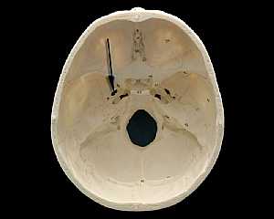
Skull Anatomy
19/10/2009 03:02:24 م
In This Section you will find detailed different Photos and images about the anatomy of the Skull bone including its surface , attachments related structures many more Items about the Skull anatomy... More Details
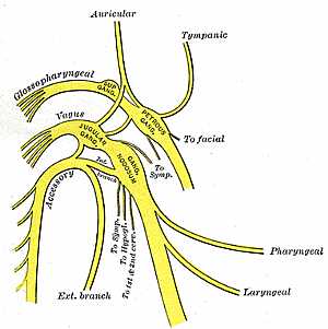
Cranial nerves IX , X and XI anatomy
30/10/2009 03:51:00 م
this image shows the cranial nerves IX,X,XI (glossopharyngeal ,vagus and accessory nerves) showing: 1. glossopharyngeal nerve 2. vagus nerve 3. accessory nerve 4. external branch 5. laryngeal nerve 6... More Details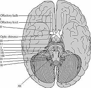
cranial nerves anatomy
14/10/2009 04:17:00 ص
this image is an inferior view of the brain showing the cranial nerves showing: 1. olfactory nerve 2. optic nerve 3. occulomoto rnerve 4. trochlear nerve 5. trigeminal nerve 6. abducent nerve 7. fasci... More Details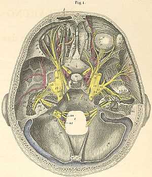
cranial nerves course in the skull
10/10/2009 03:09:00 م
this image shows the course of the cranial nerves inside the skull a) Frontal bone. b) Frontal sinus. c) Internal frontal spine. d) Foramen caecum. e) Crista galli. f) Frontal bone, orbital portion. g... More DetailsGlossopharybgeal nerve anatomy
29/10/2009 03:23:00 م
this is an image of the lateral side of the neck displaying the left glossopharyngeal nerve related to the surrounding structures showing: 1. left glossopharyngeal nerve 2. external auditory meatus 3... More Details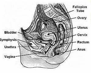
Vagina anatomy
13/11/2009 06:27:00 ص
this image shows the vagina of the female pelvis in relation to the surrounding organs this is a side view of a vertically sectioned pelvis showing: 1. fallopian tube 2. ovary 3. uterus 4. cervix 5. r... More Detailsorthopaedic joint assessment centr dr mcmahon
, , , , , , ,anatomi ligamen panggul wanita
, ,abdomen sans preparation normale
, , , , , ,world conferences on urine therapy
, , , , , , , , , ,© Copyright 2001-2022 eDoctorOnline.com