Brain MRI - Frontal
This image shows a transverse section of the brain at the level of the Frontal lobes using MRI technique (notice that the bone here is black in contrast to CT in which the bone is white)This technique shows the details of thee soft tissue like the brain in this image
This image shows the Gyri and Sulci of the brain at this level ( the gyrus is an elevation or a loop in the brain tissue and the sulcus is the groove in the brain tissue in between these gyrii)
Showing:
1. Superior Frontal Gyrus
2. Middle Frontal Gyrus
3. Inferior Frontal Gyrus
4. Central Sulcus
5. Supra-marginal Gyrus
6. Intra-parietal Sulcus
7. Angular Sulcus
8. Anterior cerebral artery

Rate Photo:


6 Ratings
Views: 7481


Link this photo to your website:
Copy the above code and paste it into your webpage, blog or forum
larynx
humerus
corpus callosum
midbrain
neck anatomy
humerus
nasal cavity
heart anatomy
midbrain
cranial nerves
eye anatomy
ear anatomy
neck muscles
muscular system
stomach anatomy
stomach
humerus bone
NOSE ANATOMY
Cerebrum
cerebral cortex
FEMUR
anatomy of the neck
humerus bone
eye anatomy
stomach anatomy
pelvic girdle
temporal lobe
Pituitary gland
visual pathway
eye diagram
eye anatomy
femur
falx cerebri
ear anatomy
vagus nerve
neck anatomy
muscular system
heart anatomy
anatomy of neck
eye diagram
lung anatomy
eye anatomy
lung anatomy
nasal cavity
pelvic girdle
visual pathway
brain anatomy
neck anatomy
skeletal system
midbrain
Most Viewed
Most Downloads
 Nose anatomy
 Humerus bone
 Eye anatomy
 Coronary arteries anatomy
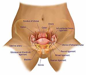 Female pelvic anatomy
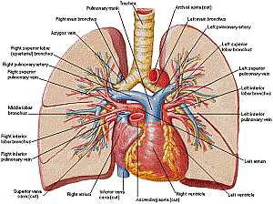 Heart and lung anatomy
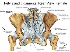 Bones and ligaments of the FEMALE Pelvis
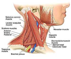 Neck Anatomy
 MidBrain anatomy
 Oral Cavity
 Stomach anatomy
 Lung anatomy
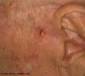 Basal Cell Carcinoma ("Rodent Ulcer" Type)
 Basal Cell Carcinoma ("Rodent Ulcer" Type)
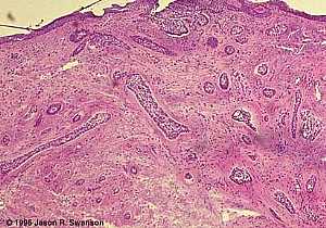 Basal Cell Carcinoma (Histology-Morpheaform Type)
 Basal Cell Carcinoma (Histology-Morpheaform Type)
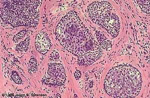 Basal Cell Carcinoma (Histology-Nodular Type - High power)
 Basal Cell Carcinoma (Histology-Nodular Type - High power)
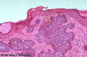 Basal Cell Carcinoma (Histology-Nodular Type- High power)
 Basal Cell Carcinoma (Histology-Nodular Type- High power)
 Skin
Skin
 Skin
Skin Nervous System -- Basic
 Nervous System -- Basic
 Brain anatomy
 Brain anatomy
 Brain anatomy
 Brain anatomy
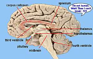 Brain anatomy
 Brain anatomy
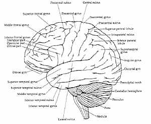 Brain anatomy
 Brain anatomy
 Head anatomy
 Head anatomy
 Brain anatomy
 Brain anatomy
eDoctorOnline.com does not provide medical advice, diagnosis or treatment.
© Copyright 2001-2022 eDoctorOnline.com
© Copyright 2001-2022 eDoctorOnline.com
Be the first one to comment on this photo!