anatomy of brain stem
midbrain, diencephalon, midbrain anatomy, mid brain, brain anatomy, lateral view of the brain, the midbrain, midbrain location, brain stem, brain stem anatomy, brain anatomy, transverse fissure, Medulla oblongata location, Brain Stem, diencephalon, calcarine fissure, diencephalon anatomy, brain stem anatomy, transverse fissure of brain, Brain Stem Anatomy,
The following are the result pages for the searched keyowrd: anatomy of brain stem
Brain anatomy
13/10/2009 12:46:00 ص
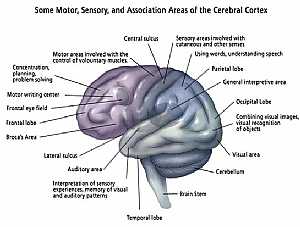
this image shows the main centers of the brain (every center has its own specific function) showing: 1. temporal lobe 2. auditory area 3. lateral sulcus 4. broca's area 5. frontal lobe 6. frontal ... More Details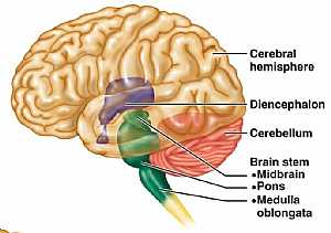
Brain anatomy
13/10/2009 12:39:00 ص
this is lateral view of the brain showing the cerebrum, cerebellum and their relation to the diencephalon (hypothalamus) and the brain stem (the root of the brain) showing: 1. cerebral hemisphere 2. d... More Details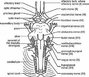
cranial nerves anatomy
14/10/2009 04:16:00 ص
this diagram shows the 12 cranial nerves and where they exactly exit from the brain showing: 1. all cranial nerves 2. olfactory bulb 3. optic chisma 4. pituitary gland 5. optic tract 6. mammillary bod... More Details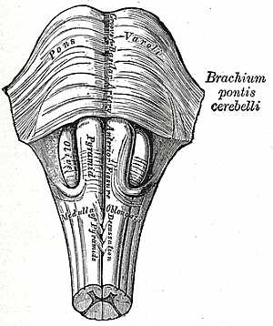
Brain stem anatomy
22/10/2009 01:50:57 م
In This Section you will find detailed different Photos and images about the anatomy of the Brain Stem including its surface , parts , related structures , midbrain anatomy , pons anatomy , medulla an... More Details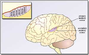
Cerebrum anatomy
22/10/2009 01:50:07 م
In This Section you will find detailed different Photos and images about the anatomy of the Cerebrum including its surface , parts , related structures , Functional areas of the brain , different cent... More Details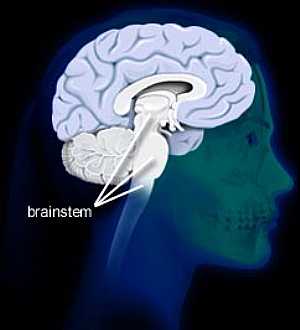
Brain stem anatomy
16/10/2009 02:00:00 م
this image shows the relation between the brain stem and the cerebri... More Details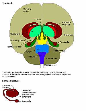
brain stem anatomy
23/10/2009 03:14:00 م
this is a anterior view of the whole brain showing the relation of the different parts of the brain stem to the rest of the brain showing: 1. cerebrum 2. fornix 3. thalamus 4. globus pallidus 5. mamm... More DetailsBrain stem anatomy
24/10/2009 03:14:00 م
this image shows the brain stem and the cerebellum and the spinal cord in relation to each other showing: 1. cerebellum 2. pons 3. midbrain 4. medulla 5. spinal cord 6. decussation of the pyramids 7.... More Details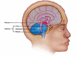
MidBrain anatomy
16/10/2009 01:59:00 م
this is image shows the location of the midbrain and the hind brain showing: 1. midbrain 2. Pons 3. Cerebellum 4. Medulla oblongata... More Details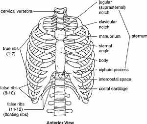
Thoracic cage anatomy
09/11/2009 03:42:00 م
this is an anterior view of the thoracic cage (the bones that forms the thorax) showing: 1. cervical vertebrae 2. jugular (suprasternal) notch 3. clavicular notch 4. manubrium 5. sternal angle 6. bod... More Details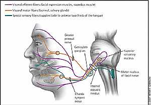
Fascial Nerve anatomy
29/10/2009 04:03:00 م
this image displays the fascial nerve branches in the face and colored according to its function showing: 1. superior salivary nucleus 2. motor nucleus of fascial nerve 3. internal acoustic meatus 4.... More DetailsRelated Searches
loading...
loading...
Top Health & Medical Articles
New Articles
Most Read
آخر كلمات البحث
orthopaedic joint assessment centr dr mcmahon
, , , , , , ,anatomi ligamen panggul wanita
, ,abdomen sans preparation normale
, , , , , ,world conferences on urine therapy
, , , , , , , , , ,eDoctorOnline.com does not provide medical advice, diagnosis or treatment.
© Copyright 2001-2022 eDoctorOnline.com
© Copyright 2001-2022 eDoctorOnline.com