anatomy pictures
humerus, heart anatomy, ear anatomy, humerus bone, eye anatomy, ear anatomy, heart anatomy, anatomy of heart, stomach, pelvic anatomy, breast anatomy, stomach anatomy, trabeculae carneae, spinal cord anatomy, pectinate muscles, anatomy of ear, skull anatomy, fossa ovalis, anatomy, heart,
The following are the result pages for the searched keyowrd: anatomy pictures
Heart anatomy
16/07/2010 05:10:26 ص
In This Section you will find detailed different Photos and images about the anatomy of the Heart including its surface , parts , related structures , Blood circulation of the heart , coronaries anato... More Details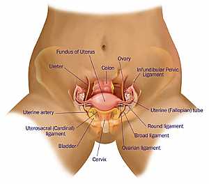
Female pelvic anatomy
13/11/2009 07:04:00 ص
this is an anterior view of the female reproductive system in the pelvic region showing: 1. fundus of the uterus 2. ureter 3. colon 4. ovary 5. infundibular pelvic ligament 6. uterine (fallopian ) tu... More Details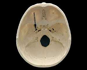
Skull Anatomy
19/10/2009 03:02:24 م
In This Section you will find detailed different Photos and images about the anatomy of the Skull bone including its surface , attachments related structures many more Items about the Skull anatomy... More Details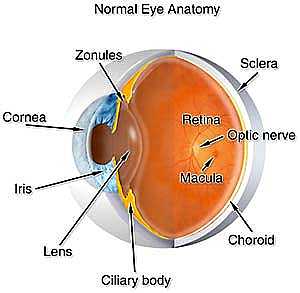
Eye anatomy
11/10/2009 03:47:00 م
this is a 3D image of the eye showing its different parts showing: 1. zonules 2. cornea 3. iris 4. lens 5. ciliary body 6. macula 7. retina 8. choroid 9. optic nerve 10. sclera... More Details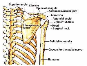
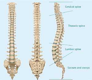
Spinal cord anatomy
22/10/2009 01:52:10 م
In This Section you will find detailed different Photos and images about the anatomy of the Spinal cord including its surface , parts , related structures , Functions of the spinal; cord , Spinal nerv... More Details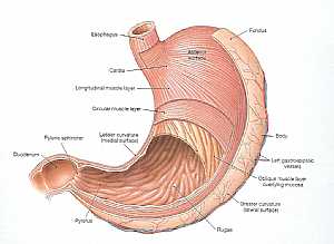
Stomach anatomy
09/12/2009 06:08:00 ص
this image shows the anatomy of the stomach showing its main features and parts.in this images we see the wall of the stomach being removed from the anterior portion to display the contents of the sto... More Details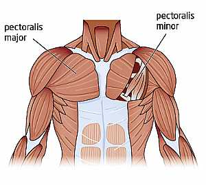
Pectoral anatomy
02/12/2009 01:33:00 م
this image shows the anatomy of the pectoral region pointing at the pectoralis major and pectoralis minor muscles (the main muscles of that region)... More Details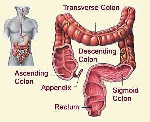
Large intestine anatomy
09/12/2009 05:53:00 ص
this image shows the anatomy of the large intestine from inside and from outside displaying its different parts and features and its relation to the rest of the body specially to the GIT system. show... More Details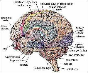
Brain anatomy
14/10/2009 04:18:00 ص
this is a detailed image for the lateral view of the cerebri and the cerebellum showing: 1. substania nigra 2. pituitary gland 3. hippocampus 4. hypothalamus 5. eye 6. basal ganglia 7. prefrontal cort... More Details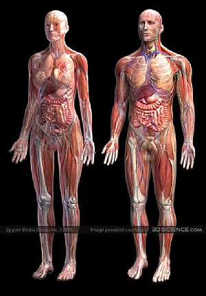
Muscles Anatomy (muscular system)
05/03/2009 02:10:00 م
this image shows the different muscles the forms and supports our human body and differs between male and female... More Details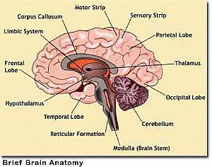
Cerebrum anatomy
13/10/2009 12:49:00 ص
this is a detailed image of the cerebrum and its parts showing: 1. temporal lobe 2. hypothalamus 3. frontal lobe 4. limbic system 5. corpus callosum 6. motor strip 7. sensory strip 8. parietal lobe 9.... More Details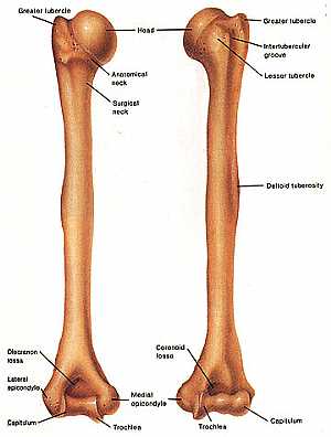
Humerus bone
04/12/2009 06:27:00 ص
this image shows the humerus bone (the bone of the arm " links the shoulder to the elbow joint") from anterior and posterior view showing: 1. greater tubercle 2. head of the humerus 3. anatom... More Details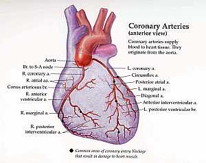
Coronary arteries anatomy
20/12/2009 02:25:00 م
this image shows the coronary arteries of the heart ( the arteries that supply the heart muscle with oxygen and nutrients) From anterior view .these arteries when occluded paretially or completely it ... More Details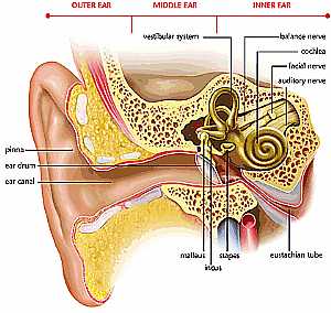
Ear anatomy
12/10/2009 04:21:00 ص
this image shows the different structures of the ear (external , middle and inner ear ) showing: 1. ear pinna 2. ear drum 3. ear canal 4. malleus 5. incus 6. stapes 7. eustacian tube 8. vestibular sy... More Details
Neck Anatomy
19/10/2009 03:05:59 م
In This Section you will find detailed different Photos and images about the anatomy of the Neck including its surface , attachments , structures , Neck arteries , Neck Veins , trachea , Esophagus an... More Details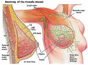
Breast anatomy
02/12/2009 01:38:00 م
this image shows the anatomy of the female breast from anterior view (on the right) and a side view of a sectioned breast (on the left) showing: 1. pectoralis major muscle 2. lymphatic drainage 3. ni... More Details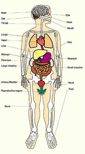
Ear anatomy
12/10/2009 04:21:00 ص
this image shows the different structures of the ear (external , middle and inner ear ) showing: 1. ear pinna 2. ear drum 3. ear canal 4. malleus 5. incus 6. stapes 7. eustacian tube 8. vestibular sy... More Details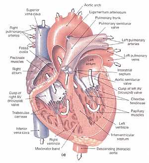
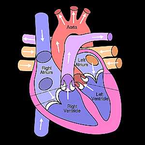
Heart anatomy
09/11/2009 03:55:00 م
this image shows the detailed section of the heart displaying all its internal parts and the vessels going into and getting out of the heart showing: 1. aortic arch 2. ligamentum arteriosum 3. pulmon... More Details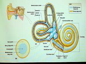
inner ear anatomy
12/10/2009 04:19:00 ص
this is a detailed image for the inner ear showing: 1. semicircular canals 2. semicircular ducts ( ant , post and middle) 3. vestibule 4. ampullae 5. maculae 6. endolymphatic sac 7. utricle 8. saccule... More Details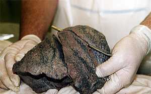
Lung anatomy
07/12/2009 06:31:00 ص
this image shows the anatomy of the right lung displaying its two main fissures the horizontal and the oblique one.they divide the right lung into three lobs "the upper lobe, the middle or the lat... More Detailsorthopaedic joint assessment centr dr mcmahon
, , , , , , ,anatomi ligamen panggul wanita
, ,abdomen sans preparation normale
, , , , , ,world conferences on urine therapy
, , , , , , , , , ,© Copyright 2001-2022 eDoctorOnline.com