coronary artery anatomy
heart anatomy, coronary arteries, coronary artery anatomy, liver anatomy, CORONARY ARTERIES ANATOMY, coronary artery, heart, anatomy of the heart, coronary artery anatomy, anatomy, anatomy of heart, anatomy of liver, coronary anatomy, coronary arteries, anatomy of the liver, image, heart valves, coronary arteries anatomy, coronary arteries, heart anatomy,
The following are the result pages for the searched keyowrd: coronary artery anatomy
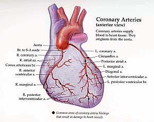
Coronary arteries anatomy
20/12/2009 02:25:00 م
this image shows the coronary arteries of the heart ( the arteries that supply the heart muscle with oxygen and nutrients) From anterior view .these arteries when occluded paretially or completely it ... More DetailsCoronary arteries anatomy
20/12/2009 02:25:00 م
this image shows the coronary arteries of the heart ( the arteries that supply the heart muscle with oxygen and nutrients) From anterior view .these arteries when occluded paretially or completely it ... More Details
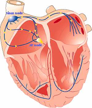
Natural pacemaker of the heart
20/12/2009 02:19:00 م
this image shows the natural passage of the nerve impulses in the cardiac muscle.This image shows the natural pacemaker of the heart (SA node)"sinoatrial node" that sinus has its own rhythmic ... More Details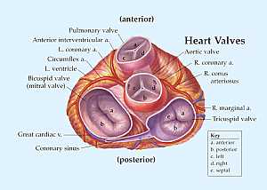
heart valve anatomy
07/01/2010 02:27:00 م
This image is a horizontal cut section of the heart at the level of the heart valves showing the four main valves of the heart with the related structures and vessels showing: 1. Pulmonary valve 2. A... More Details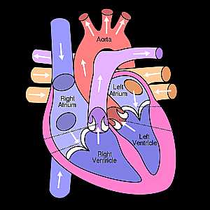
Heart anatomy
16/07/2010 05:10:26 ص
In This Section you will find detailed different Photos and images about the anatomy of the Heart including its surface , parts , related structures , Blood circulation of the heart , coronaries anato... More Details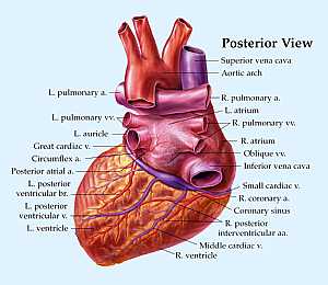
Heart anatomy
07/01/2010 02:23:00 م
This image shows the anatomy of the heart from external posterior view showing the different parts and features of the heart with the related vessels showing: 1. Superior vena cava 2. Aortic arch 3. ... More Details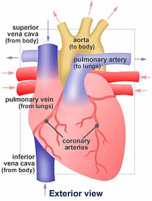
coronary arteries
20/12/2009 02:51:00 م
This image shows the anatomy of the vessels of the heart emerging from it and entering it.It also displays the circulation of the blood in these vessels. showing: 1. Superior vena cava 2. Aorta 3. Pu... More DetailsHeart anatomy
07/01/2010 02:23:00 م
This image shows the anatomy of the heart from external posterior view showing the different parts and features of the heart with the related vessels showing: 1. Superior vena cava 2. Aortic arch 3. ... More Details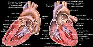
Gross anatomy of the heart
05/03/2009 04:32:00 م
this image shows the gross anatomy of a vertically sectioned heart showing: 1. brachiocephalic artery 2. left common carotid artery 3. left subclavian artery 4. left brachiocephalic vein 5. aortic ar... More DetailsHeart anatomy
07/01/2010 02:23:00 م
This image shows the anatomy of the heart from external posterior view showing the different parts and features of the heart with the related vessels showing: 1. Superior vena cava 2. Aortic arch 3. ... More Details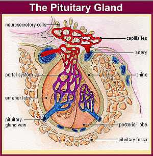
Pituitary gland anatomy
23/10/2009 03:31:00 م
this is a detailed image of the pituitary gland showing: 1. neurosecretory cells 2. portal system 3. ant. lobe 4. pituitary gland vein 5. pituitary fossa 6. post. lobe 7. artery 8. capillaries... More Details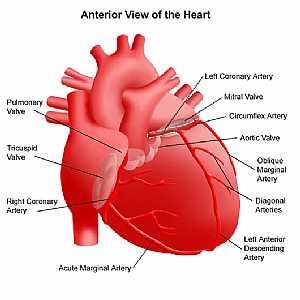
Heart anatomy
20/12/2009 02:41:00 م
this image shows the anatomy of the heart from anterior view displaying its different parts and features.we can also see the coronary arteries on the anterior aspect supplying the heart with oxygen an... More Details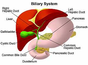
Anatomy of the liver"biliary system"
05/03/2009 04:40:00 م
this image shows the biliary system ( with the liver and the stomach) showing: 1. right hepatic duct 2. liver 3. gall bladder 4. cystic duct 5. common bile duct 6. duodenum 7. pancreatic duct 8. comm... More Details
orthopaedic joint assessment centr dr mcmahon
, , , , , , ,anatomi ligamen panggul wanita
, ,abdomen sans preparation normale
, , , , , ,world conferences on urine therapy
, , , , , , , , , ,© Copyright 2001-2022 eDoctorOnline.com