cranial nerve 9
cranial nerves, stylomastoid foramen, cranial nerves, trigeminal nerve, auriculotemporal nerve, trigeminal nerve, trigeminal nerve, cranial nerves, cranial nerve, maxillary nerve, trigeminal nerve branches, maxillary nerve, trigeminal nerve, cranial nerves, trigeminal nerve anatomy, cranial nerves anatomy, nerves, stylomastoid foramen, trigeminal nerve anatomy, zygomatic nerve,
The following are the result pages for the searched keyowrd: cranial nerve 9
Skull Anatomy
19/10/2009 03:02:24 م
In This Section you will find detailed different Photos and images about the anatomy of the Skull bone including its surface , attachments related structures many more Items about the Skull anatomy... More Details
Cranial nerves anatomy
22/10/2009 01:51:53 م
In This Section you will find detailed different Photos and images about the anatomy of the Cranial Nerves including Their types , Fascial nerve anatomy , trigeminal nerve anatomy , vagus nerve anatom... More Details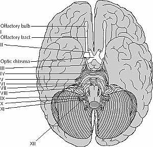
cranial nerves anatomy
14/10/2009 04:17:00 ص
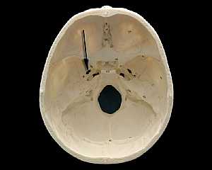
this image is an inferior view of the brain showing the cranial nerves showing: 1. olfactory nerve 2. optic nerve 3. occulomoto rnerve 4. trochlear nerve 5. trigeminal nerve 6. abducent nerve 7. fasci... More Details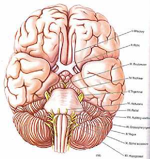
cranial nerves anatomy
14/10/2009 04:29:00 ص
a colored image for the cranial nerves exit showing: 1. olfactory nerve 2. optic nerve 3. occulomoto rnerve 4. trochlear nerve 5. trigeminal nerve 6. abducent nerve 7. fascial nerve 8. vestibulocochle... More Details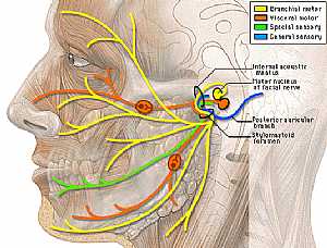
Fascial Nerve anatomy
30/10/2009 03:12:00 م
this image shows the fascial nerve in the lateral aspect of the face displaying its branches to the different muscles of the face showing: 1. internal acoustic meatus 2. posterior auricular branch 3.... More DetailsFascial Nerve anatomy
30/10/2009 03:12:00 م
this image shows the fascial nerve in the lateral aspect of the face displaying its branches to the different muscles of the face showing: 1. internal acoustic meatus 2. posterior auricular branch 3.... More Details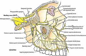
Trigeminal nerve anatomy
28/10/2009 01:55:00 ص
this images illustrates the different branches of the trigeminal nerve in the face in relation to each other [focusing on the maxillary division] showing: 1. maxillary nerve 2. meningeal branch 3. po... More DetailsCranial nerves anatomy
05/11/2009 04:03:00 ص
this image shows the all cranial nerves and displaying their effector organs showing: 1. Olfactory nerve I 2. Optic nerve II 3. Occulomotor nerve III 4. Trochlear nerve IV 5. Trigeminal nerve V 6. Ab... More Details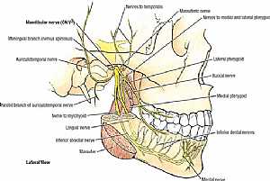
Trigeminal nerve anatomy
28/10/2009 02:07:00 ص
this images illustrates the different branches of the trigeminal nerve in the face in relation to each other [focusing on the mandibular division] showing: 1. mandibular nerve 2. meningeal branch 3. ... More Details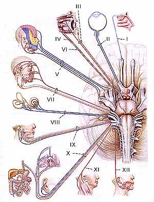
cranial nerves anatomy
14/10/2009 04:30:00 ص
this image shows each cranial nerve and its site of action ( its function)... More Details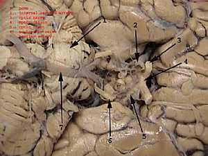
Occulomotor nerve anatomy
28/10/2009 02:10:00 ص
this image shows the oculomotor nerve at its origin in relation to the surrounding structures showing: 1. pons 2. internal carotid artery 3. optic nerve 4. optic chiasma 5. optic tract 6. occulomotor... More Details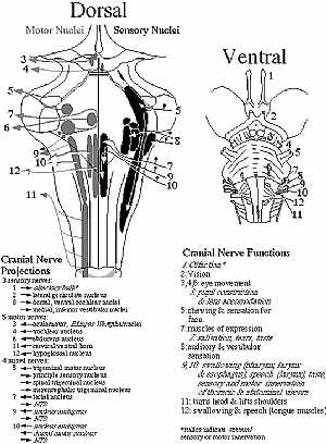
cranial nerves anatomy
14/10/2009 04:28:00 ص
this diagram shows location of the cranial nerves nuclei and their site of exit the right image is an anterior view and the left is a posterior view showing: 1. olfactory nerve 2. optic nerve 3. occul... More Details
The fascial nerve
16/10/2009 02:19:00 م
this image shows the fascial nerve (cranial nerve VII) and shows its course and the organs supplied by it showing: 1. fascial nerve in the pons 2. at stylomastoid foramen 3. cervical branch 4. tempora... More Detailscranial nerves anatomy
14/10/2009 04:17:00 ص
this image is an inferior view of the brain showing the cranial nerves showing: 1. olfactory nerve 2. optic nerve 3. occulomoto rnerve 4. trochlear nerve 5. trigeminal nerve 6. abducent nerve 7. fasci... More Details
Trigeminal nerve anatomy
28/10/2009 01:55:00 ص
this images illustrates the different branches of the trigeminal nerve in the face in relation to each other [focusing on the maxillary division] showing: 1. maxillary nerve 2. meningeal branch 3. po... More Details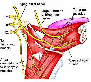
Hypoglossal nerve anatomy
03/11/2009 02:13:00 م
this image shows the cranial nerve XII "hypoglossal nerve" in the face region in the lateral aspect in relation to the surrounding structures showing: 1. hypoglossal nerve 2. lingual branch o... More Details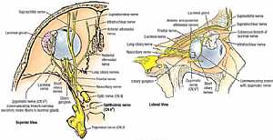
Nerve supply of the eye
28/10/2009 01:50:00 ص
this image shows the nerves supplying the eye in relation to each other from superior view (on the left) and from lateral view (on the right) showing: 1. ophthalmic nerve 2. trigeminal nerve 3. optic... More Detailsorthopaedic joint assessment centr dr mcmahon
, , , , , , ,anatomi ligamen panggul wanita
, ,abdomen sans preparation normale
, , , , , ,world conferences on urine therapy
, , , , , , , , , ,© Copyright 2001-2022 eDoctorOnline.com