face anatomy
facial nerve, anatomy of the face, geniculate ganglion, ansa cervicalis, pharyngeal tonsils, facial nerve anatomy, facial nerve anatomy, palatine tonsils, pharyngeal tonsil, palatine tonsil, stylomastoid foramen, facial nerve branches, facial nerve branches, lingual tonsils, geniculate ganglion, facial nerve branches, lingual tonsil, anatomy of the face, face anatomy, facial nerve,
The following are the result pages for the searched keyowrd: face anatomy
Anatomy of the face
10/10/2009 04:23:00 م
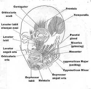
this is a detailed image for the fascial muscles ( the muscles of the face) showing: 1.frontalis m. 2. temporalis m. 3. parotid gland 4. risorius m. 5. masseter m. 6. zygomaticus major m. 7. zygomatic... More DetailsAnatomy of the face
10/10/2009 04:23:00 م
this is a detailed image for the fascial muscles ( the muscles of the face) showing: 1.frontalis m. 2. temporalis m. 3. parotid gland 4. risorius m. 5. masseter m. 6. zygomaticus major m. 7. zygomatic... More Details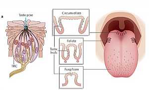
Head Anatomy
19/10/2009 03:02:56 م
In This Section you will find detailed different Photos and images about the anatomy of the Head including The Face , Muscles of the face , bones of the face and vessles many more Items about the Skul... More Details

Cranial nerves anatomy
22/10/2009 01:51:53 م
In This Section you will find detailed different Photos and images about the anatomy of the Cranial Nerves including Their types , Fascial nerve anatomy , trigeminal nerve anatomy , vagus nerve anatom... More Details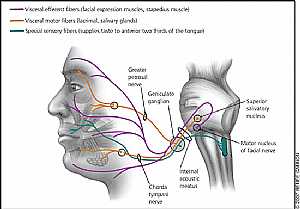
Fascial Nerve anatomy
29/10/2009 04:03:00 م
this image displays the fascial nerve branches in the face and colored according to its function showing: 1. superior salivary nucleus 2. motor nucleus of fascial nerve 3. internal acoustic meatus 4.... More Details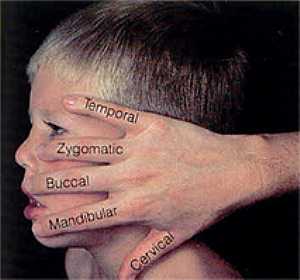
Branches of Facial nerve anatomy
30/10/2009 03:05:00 م
this image shows the branches of the facial nerve with the direction of each branch in the face... More Details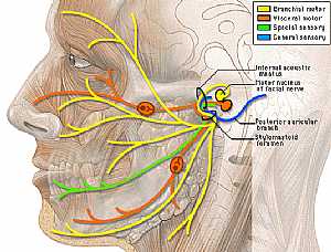
Fascial Nerve anatomy
30/10/2009 03:12:00 م
this image shows the fascial nerve in the lateral aspect of the face displaying its branches to the different muscles of the face showing: 1. internal acoustic meatus 2. posterior auricular branch 3.... More DetailsBranches of Facial nerve anatomy
30/10/2009 03:05:00 م
this image shows the branches of the facial nerve with the direction of each branch in the face... More Details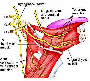
Hypoglossal nerve anatomy
03/11/2009 02:13:00 م
this image shows the cranial nerve XII "hypoglossal nerve" in the face region in the lateral aspect in relation to the surrounding structures showing: 1. hypoglossal nerve 2. lingual branch o... More Details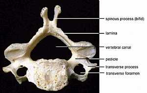
Vertebral Anatomy
19/10/2009 03:02:39 م
In This Section you will find detailed different Photos and images about the anatomy of the Vertebrae bones including their types , their surface , attachments related structures many more Items about... More Details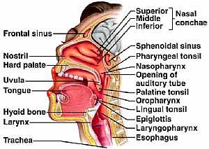
Head anatomy
12/10/2009 04:32:00 ص
this section details the nose,mouth ,the pharynx showing: 1. sup. , middle and inf. nasal conchae 2. sphenoid sinus 3. pharyngeal tonsil 4. nasopharynx 5. opening of auditory tube 6. palatine tonsil 7... More Details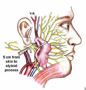
Fascial nerve anatomy
29/10/2009 03:33:00 م
tbis image shows the surface anatomy of the point of exit of the fascial nerve from the intracranial cavity to the face... More DetailsFascial nerve anatomy
29/10/2009 03:33:00 م
tbis image shows the surface anatomy of the point of exit of the fascial nerve from the intracranial cavity to the face... More Detailsorthopaedic joint assessment centr dr mcmahon
, , , , , , ,anatomi ligamen panggul wanita
, ,abdomen sans preparation normale
, , , , , ,world conferences on urine therapy
, , , , , , , , , ,© Copyright 2001-2022 eDoctorOnline.com