ischial spine
ilium bone, coxal bone, pelvic anatomy, anterior superior iliac spine, pelvis, PUDENDAL NERVE, sacrotuberous ligament, obturator foramen, sacrum, Pelvis, linea terminalis, iliac bone, female pelvic anatomy, pudendal canal, lesser sciatic foramen, pudendal nerve, iliac spine, pudendal nerve anatomy, ischium bone, pelvis anatomy,
The following are the result pages for the searched keyowrd: ischial spine
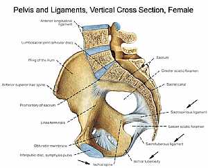
Bones and ligaments of the FEMALE Pelvis
22/11/2009 06:07:00 ص
this image is a longitudinal cross section in the female pelvic brim (the bones and ligaments of the female pelvic region) displaying a lateral view of that sectioned pelvis showing: 1. anterior long... More DetailsBones and ligaments of the FEMALE Pelvis
22/11/2009 06:07:00 ص
this image is a longitudinal cross section in the female pelvic brim (the bones and ligaments of the female pelvic region) displaying a lateral view of that sectioned pelvis showing: 1. anterior long... More Details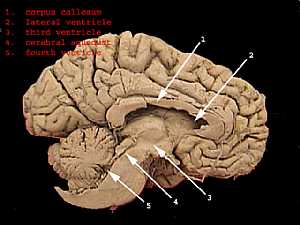
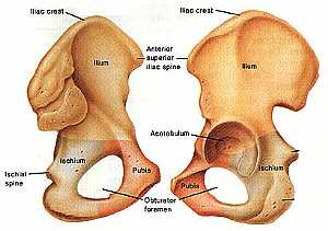
Hib bone anatomy
04/12/2009 07:22:00 ص
this image shows the anatomy of the hib bone from anterio-lateral (on the left) and from posterio-lateral view(on the right) showing: 1. iliac crest 2. ilium bone 3. ischial bone 4. pubis 5. obturato... More Details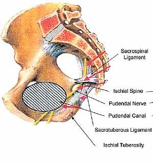
Pudendal nerve anatomy
13/11/2009 06:43:00 ص
this image shows the pudendal nerve in the pelvic showing its course ond relation to the surrounding structures showing: 1. sacrospinal ligament 2. iscial spine 3. pudendal nerve 4. pudendal canal 5.... More Details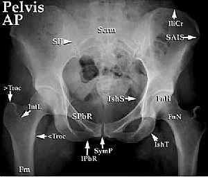
Normal Pelvis X-Ray
09/01/2012 12:34:00 م
This is a X-Ray Image of Pelvis Antero-Posterior View,Showing the pelvis from the front. Showing : 1. 2 Iliac Crests (IliCr) 2. Greater Trochanter (>Troc) 3. Intertrochanteric Line (IntL) 4. Lesser T... More Details
Pelvis Anatomy
06/11/2009 01:06:01 م
In This Section you will find detailed different Sections about the different organs and structures in the region of the Pelvis including Male pelvis anatomy , Female pelvis anatomy , pelvic girdle an... More Details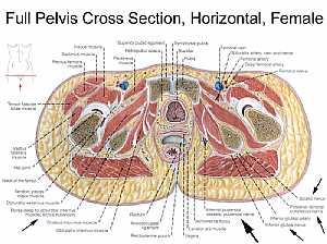
Female pelvic anatomy
13/11/2009 07:31:00 ص
this image is a horizontal section in a female pelvis shows the different pelvic organs and structures in relation to each other showing: 1. iliacus muscle 2. sartorius muscle 3. rectus femoris muscl... More Details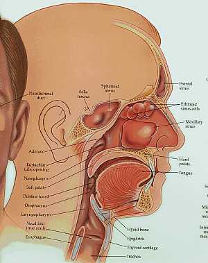
anatomy of the head and neck
09/10/2009 03:44:00 م
this image shows longitudinal section in the head and neck with their details (the frontal,nasal,oral and neck regions) showing: 1. nasolacrimal duct 2. sella turcica 3. sphenoid sinus 4. frontal sinu... More DetailsRelated Searches
loading...
loading...
Top Health & Medical Articles
New Articles
Most Read
آخر كلمات البحث
orthopaedic joint assessment centr dr mcmahon
, , , , , , ,anatomi ligamen panggul wanita
, ,abdomen sans preparation normale
, , , , , ,world conferences on urine therapy
, , , , , , , , , ,eDoctorOnline.com does not provide medical advice, diagnosis or treatment.
© Copyright 2001-2022 eDoctorOnline.com
© Copyright 2001-2022 eDoctorOnline.com