mammillary body
third ventricle, lateral ventricle, fourth ventricle, 4th ventricle, brain ventricles, ventricles of the brain, lateral ventricles, 3rd ventricle, lateral ventricle of brain, third ventricle of the brain, 3rd ventricle of brain, brain stem anatomy, third ventricle brain, fourth ventricle of the brain, hypothalamus and pituitary gland, ventricles of brain, image, third ventricle of brain, mammillary body, brain stem,
The following are the result pages for the searched keyowrd: mammillary body
cranial nerves anatomy
14/10/2009 04:16:00 ص
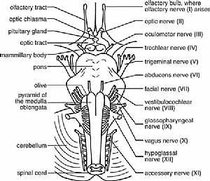
this diagram shows the 12 cranial nerves and where they exactly exit from the brain showing: 1. all cranial nerves 2. olfactory bulb 3. optic chisma 4. pituitary gland 5. optic tract 6. mammillary bod... More Detailscranial nerves anatomy
14/10/2009 04:16:00 ص
this diagram shows the 12 cranial nerves and where they exactly exit from the brain showing: 1. all cranial nerves 2. olfactory bulb 3. optic chisma 4. pituitary gland 5. optic tract 6. mammillary bod... More Details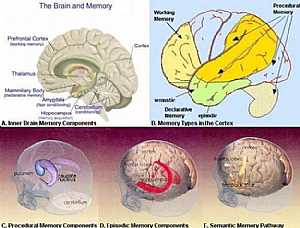
Memory centers anatomy
03/11/2009 02:35:00 م
this image shows the memory centers in the cerebral cortex( the centers responsible for our memory) showing: 1. prefrontal cortex"working memory" 2. thalamus 3. mammillary body "declarati... More Details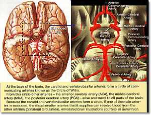
Circulus of willis
13/10/2009 01:24:00 ص
this image shows what is called circulus of wiilis arteries that are connected together in the form of a circle placed just under the two cerbri just above the pituitary gland showing: 1. internal car... More Details
Cranial nerves anatomy
22/10/2009 01:51:53 م
In This Section you will find detailed different Photos and images about the anatomy of the Cranial Nerves including Their types , Fascial nerve anatomy , trigeminal nerve anatomy , vagus nerve anatom... More DetailsMemory centers anatomy
03/11/2009 02:35:00 م
this image shows the memory centers in the cerebral cortex( the centers responsible for our memory) showing: 1. prefrontal cortex"working memory" 2. thalamus 3. mammillary body "declarati... More Details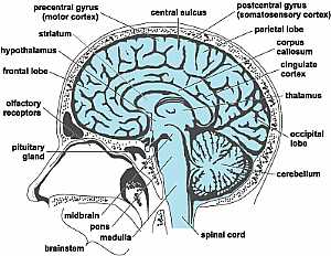
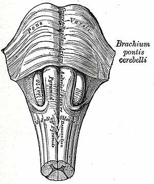
Brain stem anatomy
22/10/2009 01:50:57 م
In This Section you will find detailed different Photos and images about the anatomy of the Brain Stem including its surface , parts , related structures , midbrain anatomy , pons anatomy , medulla an... More Details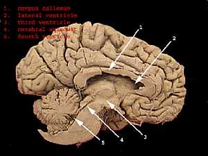
Brain ventricles
13/10/2009 01:20:00 ص
this section that shows the medial wall of the cerebral hemisphere shows the main four ventricles of the brain showing: 1. corpus callosum 2. lateral ventricle 3. cerebral aquiduct 4. fourth ventricle... More Details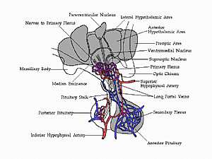
connection between hypothalamus and pituitary gland
23/10/2009 03:00:00 م
this shows the connection between the ant. pituitary and the hypothalamus showing: 1. ant. pituitary 2. lateral hypothalamic area 3. ant. hypothatlamic area 4. preoptic area 5. ventromedial nucleus 6... More Detailsorthopaedic joint assessment centr dr mcmahon
, , , , , , ,anatomi ligamen panggul wanita
, ,abdomen sans preparation normale
, , , , , ,world conferences on urine therapy
, , , , , , , , , ,© Copyright 2001-2022 eDoctorOnline.com