middle meningeal artery course
facial canal, vidian nerve, otic ganglion, middle meningeal artery, meningeal artery, deep petrosal nerve, auriculotemporal nerve, sphenopalatine ganglion, sphenopalatine foramen, foramen rotundum, maxillary nerve, posterior meningeal artery, images of branches of auriculotemporal nerve, lingual nerve branch from, vidian canal, middle meningeal artery course, arterial supply to skull, great petrosal nerve, otic ganglion pathway, auricular branch of vagus nerve,
The following are the result pages for the searched keyowrd: middle meningeal artery course
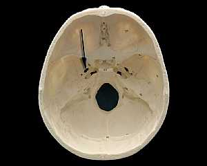
Skull Anatomy
19/10/2009 03:02:24 م
In This Section you will find detailed different Photos and images about the anatomy of the Skull bone including its surface , attachments related structures many more Items about the Skull anatomy... More Details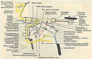
Fascial Nerve anatomy
01/11/2009 02:38:00 م
this diagram displays the course and branches of the fascial nerve showing: 1. fascial nerve nucleus 2. internal auditory meatus 3. small superfascial petrosal nerve 4. tympanic plexus 5. chorda tymp... More DetailsRelated Searches
loading...
loading...
Top Health & Medical Articles
New Articles
Most Read
آخر كلمات البحث
orthopaedic joint assessment centr dr mcmahon
, , , , , , ,anatomi ligamen panggul wanita
, ,abdomen sans preparation normale
, , , , , ,world conferences on urine therapy
, , , , , , , , , ,eDoctorOnline.com does not provide medical advice, diagnosis or treatment.
© Copyright 2001-2022 eDoctorOnline.com
© Copyright 2001-2022 eDoctorOnline.com