optic foramen
foramen rotundum, hypoglossal canal, jugular foramen, inferior orbital fissure, superior orbital fissure, foramen ovale skull, whitnall s tubercle, optic canal, optic foramen, foramen rotundum, lacrimal fossa, jugular foramen, carotid canal, orbital cavity, whitnall tubercle, foramen lacerum, hypoglossal canal, foramen rotundum, superior orbital fissure, whitnall s tubercle,
The following are the result pages for the searched keyowrd: optic foramen
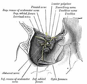
Orbital cavity anatomy
29/10/2009 03:27:00 م
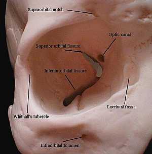
this image shows the orbital cavity detailing its different parts and structures showing: 1. supraorbital notch 2. superior orbital fissure 3. inferior orbital fissure 4. optic foramen 5. lacrimal fo... More Details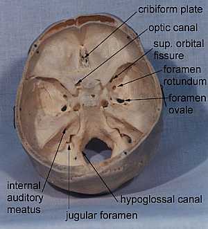
the main openings in the skull
10/10/2009 03:33:00 م
FORAMEN : CN cribiform plate I optic canal II sup. orbital fissure III, IV, Va, VI foramen rotundum Vb foramen ovale Vc int. auditory meatus VII, VIII jugular foramen IX, X, XI hypoglossal ca... More DetailsOrbital cavity anatomy
29/10/2009 03:27:00 م
this image shows the orbital cavity detailing its different parts and structures showing: 1. supraorbital notch 2. superior orbital fissure 3. inferior orbital fissure 4. optic foramen 5. lacrimal fo... More Details
Eye Anatomy
19/10/2009 03:05:30 م
In This Section you will find detailed different Photos and images about the anatomy of the Ear including its surface , attachments related structures , eye muscles , eye ball and many more Items abo... More Detailsthe main openings in the skull
10/10/2009 03:33:00 م
FORAMEN : CN cribiform plate I optic canal II sup. orbital fissure III, IV, Va, VI foramen rotundum Vb foramen ovale Vc int. auditory meatus VII, VIII jugular foramen IX, X, XI hypoglossal ca... More Details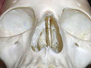
Skull Anatomy
10/10/2009 04:24:00 م
this is an anterior view of the skull showing two orbital cavities above and the nasal cavity below... More Details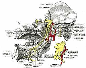
Nerve supply of the eye 3
25/10/2009 04:12:00 م
this image shows the nerves supplying the eye at the apex of the orbital cavity of the skull in relation to each other and to the other strucures there showing: 1. optic foramen 2. inf. orbital fissu... More Details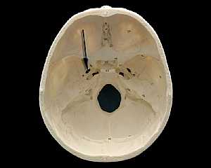
Skull Anatomy
19/10/2009 03:02:24 م
In This Section you will find detailed different Photos and images about the anatomy of the Skull bone including its surface , attachments related structures many more Items about the Skull anatomy... More Details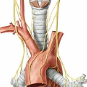
Vagus nerve anatomy
29/10/2009 03:25:00 م
this image shows the vagus nerve (right and left) in relation to the aortic arch at the left bronchus... More Details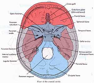
Base of the skull
10/10/2009 04:25:00 م
this a detailed image for the floor of the cranial cavity showing: 1. groove for superior sagital sinus 2. groove for ant. meningeal vessels 3. foramen caecum 4. crista gali 5. slit for nasociliary ne... More DetailsRelated Searches
loading...
loading...
Top Health & Medical Articles
New Articles
Most Read
آخر كلمات البحث
orthopaedic joint assessment centr dr mcmahon
, , , , , , ,anatomi ligamen panggul wanita
, ,abdomen sans preparation normale
, , , , , ,world conferences on urine therapy
, , , , , , , , , ,eDoctorOnline.com does not provide medical advice, diagnosis or treatment.
© Copyright 2001-2022 eDoctorOnline.com
© Copyright 2001-2022 eDoctorOnline.com