patellar tendon
lower limb anatomy, femur anatomy, which type of section separates the kneecap from the lower limb, which type of section separates the knee cap from the lower limb, infrapatellar synovial fold, infrapatellar bursa anatomy, medial condyle, lateral patellar retinaculum, infrapatellar retinaculum, horizontal setion of the lower limb, fibular nerve, lateral and medial parapatellar retinaculum, PoplitealMuscle Of Femur, lateral retinaculum of knee anatomy, bones ligaments of knee, patellar tendon, biceps femoris, medial and lateral anterial lower leg basil cell carcinoma, knee anatomy, section that separates knee cap from lower limb,
The following are the result pages for the searched keyowrd: patellar tendon
Lower limb anatomy
01/12/2009 06:54:00 ص
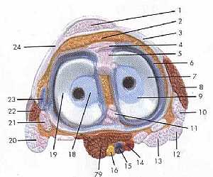
this image shows the knee joint from superior view displaying its contents and surroundings in relation to each other showing: 1. patellar tendon 2. adipose body of patella 3. medial patellar tendon ... More Details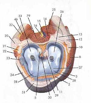
Lower limb anatomy
02/12/2009 11:25:00 ص
this image shows a horizontal section in the lower limb at the leel of the knee joint just under the femur bone displaying the muscles ,nerves , veins and arteries in relation to each other showing: ... More Detailsorthopaedic joint assessment centr dr mcmahon
, , , , , , ,anatomi ligamen panggul wanita
, ,abdomen sans preparation normale
, , , , , ,world conferences on urine therapy
, , , , , , , , , ,© Copyright 2001-2022 eDoctorOnline.com