shoulder and neck anatomy
neck anatomy, neck muscles, anatomy of the neck, neck anatomy, anatomy of neck, muscles of the neck, sternocleidomastoid, neck, neck anatomy pictures, neck anatomy, neck anatomy, neck muscles anatomy, neck anatomy, neck muscles, anatomy neck, muscles of the neck, omohyoid muscle, muscles in the neck, anatomy of the neck, anatomy of neck,
The following are the result pages for the searched keyowrd: shoulder and neck anatomy
Neck Anatomy
19/10/2009 03:05:59 م
In This Section you will find detailed different Photos and images about the anatomy of the Neck including its surface , attachments , structures , Neck arteries , Neck Veins , trachea , Esophagus an... More Details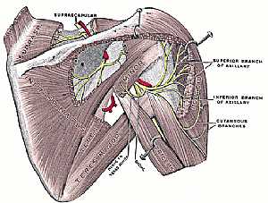
Shoulder joint anatomy
18/12/2009 07:38:00 ص
this image shows the anatomy of the shoulder joint from posterior view displaying the bones,ligaments,muscles,nerves and vessels of that region. (nerves are in yellow color,arteries are in red color) ... More Details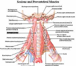
Neck Anatomy
11/10/2009 04:06:00 م
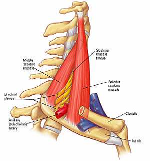
this is a back view of the muscles of the neck showing: 1. longus capitus m. 2. longus coli 3. ant. scalene m. 4. middle scalene m. 5. phrenic nerve 6. post. scalene m. 7. brachial plexus 8. subclavia... More Details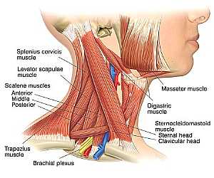
Neck Anatomy
11/10/2009 03:40:00 م
this is the muscles of the neck showing: 1. splenius cervicis m. 2. levator scapulae m. 3. masseter m. 4. digastric m. 5. sternocleidomastoid m. 6. brachial plexus 7. trapezius m. 8. ant. , middle and... More Details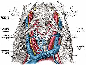
Neck anatomy
11/10/2009 04:02:00 م
this image shows the major vessels in the neck region and their relations to each other showing: 1. sternothyroid m. 2. inf. thyroid vein 3. middle thyroid vein 4. cricothyroid m. 5. sternothyroid art... More DetailsNeck Anatomy
11/10/2009 03:40:00 م
this is the muscles of the neck showing: 1. splenius cervicis m. 2. levator scapulae m. 3. masseter m. 4. digastric m. 5. sternocleidomastoid m. 6. brachial plexus 7. trapezius m. 8. ant. , middle and... More DetailsNeck Anatomy
11/10/2009 04:06:00 م
this is a back view of the muscles of the neck showing: 1. longus capitus m. 2. longus coli 3. ant. scalene m. 4. middle scalene m. 5. phrenic nerve 6. post. scalene m. 7. brachial plexus 8. subclavia... More DetailsShoulder joint anatomy
18/12/2009 07:38:00 ص
this image shows the anatomy of the shoulder joint from posterior view displaying the bones,ligaments,muscles,nerves and vessels of that region. (nerves are in yellow color,arteries are in red color) ... More Details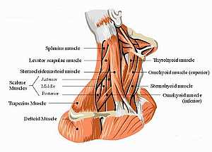
Muscles of the neck
11/10/2009 04:10:00 م
a detailed image of the muscles of the neck showing: 1. splenius m. 2. levator scapulae m. 3. sternocleidomastoid m. 4. scalene muscles 5. trapezius m. 6. deltoid m. 7. omohyoid m. 8. omohyoid muscle ... More DetailsRelated Searches
loading...
loading...
Top Health & Medical Articles
New Articles
Most Read
آخر كلمات البحث
orthopaedic joint assessment centr dr mcmahon
, , , , , , ,anatomi ligamen panggul wanita
, ,abdomen sans preparation normale
, , , , , ,world conferences on urine therapy
, , , , , , , , , ,eDoctorOnline.com does not provide medical advice, diagnosis or treatment.
© Copyright 2001-2022 eDoctorOnline.com
© Copyright 2001-2022 eDoctorOnline.com