shoulder nerves anatomy
spinal accessory nerve, spinal accessory nerve, vagus nerve, vagus nerve anatomy, ansa cervicalis, great auricular nerve, accessory nerve, vagus nerve location, greater auricular nerve, shoulder anatomy, pace maker, spinal accessory nerve anatomy, great auricular nerve, greater auricular nerve, spinal accessory nerve anatomy, image, spinal nerves, accessory nerve anatomy, geniohyoid muscle, hypoglossal nerve,
The following are the result pages for the searched keyowrd: shoulder nerves anatomy
Shoulder joint anatomy
18/12/2009 07:38:00 ص
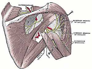
this image shows the anatomy of the shoulder joint from posterior view displaying the bones,ligaments,muscles,nerves and vessels of that region. (nerves are in yellow color,arteries are in red color) ... More Details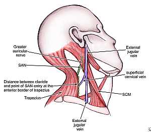
Accessory nerve anatomy
29/10/2009 03:42:00 م
this image shows the spinal part of the accessory nerve passing in the posterior triangle of the neck showing: 1. Great auricular nerve 2. Spinal accessory nerve 3. trapezius m. 4. external jugular n... More Details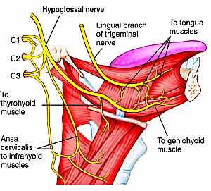
Hypoglossal nerve anatomy
03/11/2009 02:13:00 م
this image shows the cranial nerve XII "hypoglossal nerve" in the face region in the lateral aspect in relation to the surrounding structures showing: 1. hypoglossal nerve 2. lingual branch o... More Details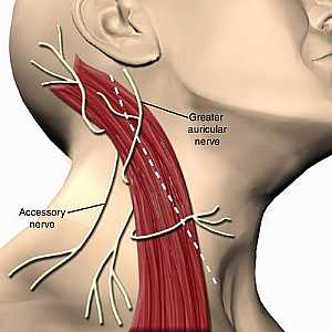
Accessory nerve anatomy
29/10/2009 03:56:00 م
this image shows the course of the accessory nerve in the neck region showing: 1. great auricular nerve 2. spinal accessory nerve 3. sternocleiomastoid muscle... More DetailsAccessory nerve anatomy
29/10/2009 03:56:00 م
this image shows the course of the accessory nerve in the neck region showing: 1. great auricular nerve 2. spinal accessory nerve 3. sternocleiomastoid muscle... More Details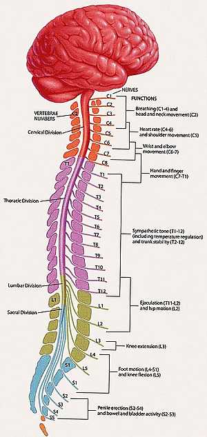
Spinal nerves anatomy
23/10/2009 03:42:00 م
this image shows the spinal cord and the emerging nerves from it showing: 1. 8 cervical spinal nerves 2. 12 thoracic spinal nerves 3. 5 lumbar spinal nerves 4. 5 sacral spinal nerves 5. 1 coccygeal s... More Details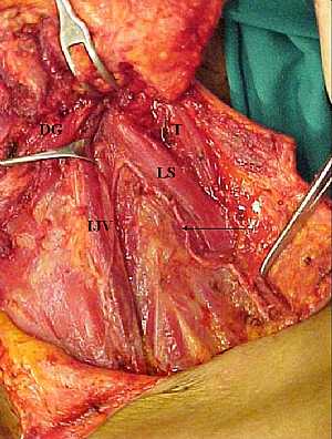
Spinal accessory nerve anatomy
29/10/2009 03:47:00 م
this image shows the spinal part of accessory nerve in the posterior triangle of the neck showing: 1. digastric muscle 2. internal jugular vein 3. accessory nerve (arrowed) 4. trapezius muscle... More Details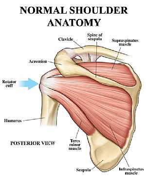
Shoulder joint anatomy
18/12/2009 07:20:00 ص
this image shows the anatomy of the shoulder joint from posterior view displaying the bones, tendons and muscles of the joint in relation to each other. showing: 1. Supraspinatus muscle 2. Spine of t... More Details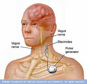
Vagus nerve anatomy
01/11/2009 02:28:00 م
this image shows the course of the vagus nerve descending from the brain to the heart ( attached to it the pulse generator "pace maker")... More DetailsRelated Searches
loading...
loading...
Top Health & Medical Articles
New Articles
Most Read
آخر كلمات البحث
orthopaedic joint assessment centr dr mcmahon
, , , , , , ,anatomi ligamen panggul wanita
, ,abdomen sans preparation normale
, , , , , ,world conferences on urine therapy
, , , , , , , , , ,eDoctorOnline.com does not provide medical advice, diagnosis or treatment.
© Copyright 2001-2022 eDoctorOnline.com
© Copyright 2001-2022 eDoctorOnline.com