sinus
nasal cavity, NOSE ANATOMY, nasal cavity, choana, nose anatomy, NASAL ANATOMY, Nasal anatomy, choana, sinus cavity, anatomy of nose, pharyngeal tonsil, pharyngeal tonsil, anatomy of nose, nasal vestibule, sphenoid sinus, anatomy of the nose, nose anatomy, anatomy of the nose, nasal vestibule, anatomi hidung,
The following are the result pages for the searched keyowrd: sinus
Nose anatomy
11/10/2009 03:55:00 م
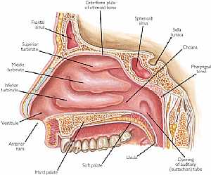
this is a longitudinal section in the nasal cavity showing its medial wall showing: 1. frontal sinus 2. superior turbinate 3. middle turbinate 4. inferior turbinate 5. vestibule 6. hard palate 7. soft... More Details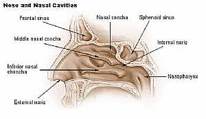
Nasal Cavity
11/10/2009 04:01:00 م
this image shows the medial wall of the nasal cavity with its elevations (choncea) showing: 1. nasal concha 2. sphenoid sinus 3. internal naris 4. nasopharynx 5. external naris 6. inf. nasal concha 7.... More DetailsNose anatomy
11/10/2009 03:55:00 م
this is a longitudinal section in the nasal cavity showing its medial wall showing: 1. frontal sinus 2. superior turbinate 3. middle turbinate 4. inferior turbinate 5. vestibule 6. hard palate 7. soft... More Details
Nose Anatomy
19/10/2009 03:05:45 م
In This Section you will find detailed different Photos and images about the anatomy of the Nose including its surface , attachments , related structures , nose bone , nose vessels and many more Item... More Details
Head Anatomy
19/10/2009 03:02:56 م
In This Section you will find detailed different Photos and images about the anatomy of the Head including The Face , Muscles of the face , bones of the face and vessles many more Items about the Skul... More Details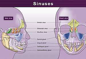
Head anatomy
11/10/2009 04:19:00 م
this shows the air sinuses of the skull ( these are spaces inside the skull bones filled with air) showing: 1. frontal sinus (yellow area) 2. ethmoidal sinus (orange are) 3. maxillary sinus (blue area... More Details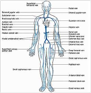
Circulation -- Venous (circulatory system)
27/04/2006
Your veins carry blood back toward the heart. Tiny vessels called capillaries in organs and tissues of the body deliver deoxygenated blood into small veins called venules, which join to form veins. Bl... More DetailsHead anatomy
11/10/2009 04:19:00 م
this shows the air sinuses of the skull ( these are spaces inside the skull bones filled with air) showing: 1. frontal sinus (yellow area) 2. ethmoidal sinus (orange are) 3. maxillary sinus (blue area... More Details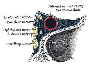
Cavernous sinus anatomy
30/10/2009 03:38:00 م
this image shows the cavernous sinus displaying the structures penetrating it showing: 1. internal carotid artery 2. oculomotor nerve 3. trochlear nerve 4. ophthalmic nerve 5. abducent nerve 6. maxil... More DetailsCavernous sinus anatomy
30/10/2009 03:38:00 م
this image shows the cavernous sinus displaying the structures penetrating it showing: 1. internal carotid artery 2. oculomotor nerve 3. trochlear nerve 4. ophthalmic nerve 5. abducent nerve 6. maxil... More DetailsRelated Searches
loading...
loading...
Top Health & Medical Articles
New Articles
Most Read
آخر كلمات البحث
orthopaedic joint assessment centr dr mcmahon
, , , , , , ,anatomi ligamen panggul wanita
, ,abdomen sans preparation normale
, , , , , ,world conferences on urine therapy
, , , , , , , , , ,eDoctorOnline.com does not provide medical advice, diagnosis or treatment.
© Copyright 2001-2022 eDoctorOnline.com
© Copyright 2001-2022 eDoctorOnline.com