spine nerve diagram
spinal accessory nerve, denticulate ligament, accessory nerve, spinal nerve, great auricular nerve, spinal accessory nerve anatomy, posterior median sulcus, image, accessory nerve anatomy, greater auricular nerve, posterior triangle of the neck, denticulate ligaments, spinal accessory nerve, external jugular anatomy, meningeal branch of spinal nerve, lateral horn, accessory nerve, spinal nerves, posterior triangle, image,
The following are the result pages for the searched keyowrd: spine nerve diagram
Spinal nerve anatomy
22/10/2009 02:37:00 م
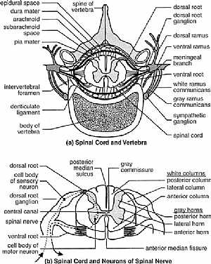
this image is a cut section in the spinal cord showing the spinal nerve emerging from it showing: (a) 1. spine of the ebidura 2. epidural space 3. dura matter 4. arachnoid space 5. subarachnoid space... More Details
Accessory nerve anatomy
29/10/2009 03:42:00 م
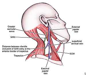
this image shows the spinal part of the accessory nerve passing in the posterior triangle of the neck showing: 1. Great auricular nerve 2. Spinal accessory nerve 3. trapezius m. 4. external jugular n... More Details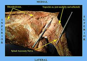
Spinal accessory nerve anatomy
29/10/2009 04:01:00 م
another image for the accessory nerve in the neck showing: 1. spinal accessory nerve 2. trapezius nerve 3. rhomboid muscle... More DetailsRelated Searches
loading...
loading...
Top Health & Medical Articles
New Articles
Most Read
آخر كلمات البحث
orthopaedic joint assessment centr dr mcmahon
, , , , , , ,anatomi ligamen panggul wanita
, ,abdomen sans preparation normale
, , , , , ,world conferences on urine therapy
, , , , , , , , , ,eDoctorOnline.com does not provide medical advice, diagnosis or treatment.
© Copyright 2001-2022 eDoctorOnline.com
© Copyright 2001-2022 eDoctorOnline.com