superior colliculus
visual pathway, superior colliculus, optic chiasm, visual pathway, auditory pathway, visual pathway, visual cortex, optic chiasm, midbrain, human organs, visual pathways, optic chiasm, optic tract, colliculus, visual pathways, optic pathway, visual pathways, superior colliculi, optic pathway,
The following are the result pages for the searched keyowrd: superior colliculus
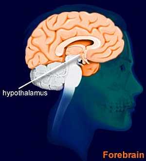
Hypothalamus anatomy
16/10/2009 02:16:00 م
this image shows the relation of the hypothalamus to the rest of the nervous system... More Details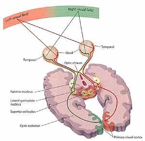
Visual pathway
03/11/2009 03:29:00 م
this image shows the visual pathway that carry sensation form the eye to the cerebral cortex showing: 1. temporal and nasal retina 2. optic nerve 3. optic chiasm 4. optic tract 5. pulvinar nucleus 6.... More DetailsVisual pathway
03/11/2009 03:29:00 م
this image shows the visual pathway that carry sensation form the eye to the cerebral cortex showing: 1. temporal and nasal retina 2. optic nerve 3. optic chiasm 4. optic tract 5. pulvinar nucleus 6.... More Details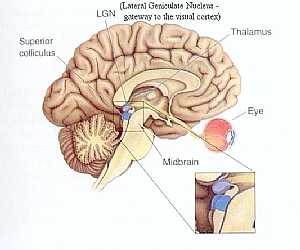
Visual pathway
16/10/2009 02:17:00 م
this image shows the tract of the visual pathway showing: 1. eye 2. midbrain 3. thalamus 4. lateral geniculate body 5. superior colliculus... More Details
Head and Neck Anatomy
09/10/2009 12:09:57 م
In This Section you will find detailed different Sections about the different organs and structures in the region of the head and neck including The skull , the vertebrae , the face , the eyes , the ... More Details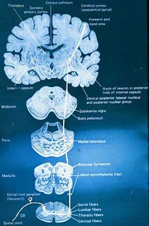
Pathways of C.N.S
22/10/2009 01:52:27 م
In This Section you will find detailed different Photos and images about the anatomy of the Pathways of the CNS including their types , spinothalamic track anatomy , ascending tracks anatomy , descend... More Details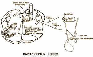
Baroreceptor reflex pathway
02/11/2009 02:20:00 م
this image shows the pathway of the baroreceptor reflex "the reflex that shares in the control of normal blood pressure" showing: 1. aortic baroreceptor 2. aortic body 3. carotid sinus 4. car... More Details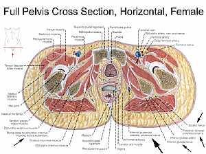
Female pelvic anatomy
13/11/2009 07:31:00 ص
this image is a horizontal section in a female pelvis shows the different pelvic organs and structures in relation to each other showing: 1. iliacus muscle 2. sartorius muscle 3. rectus femoris muscl... More Details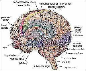
Brain anatomy
14/10/2009 04:18:00 ص
this is a detailed image for the lateral view of the cerebri and the cerebellum showing: 1. substania nigra 2. pituitary gland 3. hippocampus 4. hypothalamus 5. eye 6. basal ganglia 7. prefrontal cort... More Details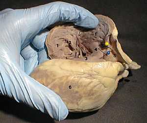
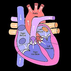
Heart anatomy
11/01/2010 03:13:00 م
This image shows a cut section in a heart from a cadaver showing some of its internal structure showing: 1. Right Ventricle 2. Papillary Muscle 3. Moderator Band 4. Pulmonary Semilunar Valve 5. Pulmo... More Details
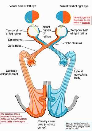
Visual pathway
28/10/2009 02:54:00 ص
this image shows the visual pathway illustrating the different fields of vision every side of the optic tract supply Note that nasal fibers from nasal retina of both sides cross at the optic chiasma ... More DetailsVisual pathway
16/10/2009 02:17:00 م
this image shows the tract of the visual pathway showing: 1. eye 2. midbrain 3. thalamus 4. lateral geniculate body 5. superior colliculus... More Details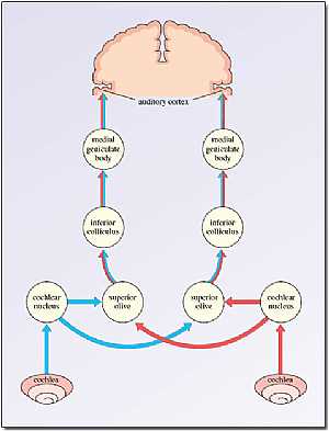
Auditory pathway
05/11/2009 03:41:00 ص
this image shows the pathway of the auditory system "the system responsible for our sense of hearing" (displays the pathway from both our left and right ears) showing: 1. cochlea "same si... More Details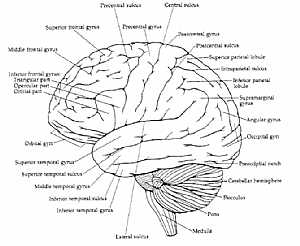
Heart anatomy
16/07/2010 05:10:26 ص
In This Section you will find detailed different Photos and images about the anatomy of the Heart including its surface , parts , related structures , Blood circulation of the heart , coronaries anato... More DetailsRelated Searches
loading...
loading...
Top Health & Medical Articles
New Articles
Most Read
آخر كلمات البحث
orthopaedic joint assessment centr dr mcmahon
, , , , , , ,anatomi ligamen panggul wanita
, ,abdomen sans preparation normale
, , , , , ,world conferences on urine therapy
, , , , , , , , , ,eDoctorOnline.com does not provide medical advice, diagnosis or treatment.
© Copyright 2001-2022 eDoctorOnline.com
© Copyright 2001-2022 eDoctorOnline.com