thyroid anatomy
neck anatomy, neck anatomy, cuneiform cartilage, cuneiform cartilage, anatomy of the neck, anatomy of neck, anatomy of the larynx, cricoid cartilage, larynx anatomy, ANATOMY OF THE LARYNX, arytenoid cartilage, anatomy neck, arytenoid cartilage, anatomy of larynx, vocal ligament, cricotracheal ligament, cricoid cartilage, cricotracheal ligament, laryngeal cartilages, Cricothyroid Ligament,
The following are the result pages for the searched keyowrd: thyroid anatomy
Anatomy of the larynx
11/10/2009 03:44:00 م
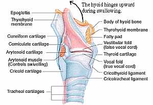
this is the structure of the larynx ( the throat area ) showing: 1. epiglottis 2. thyroid membrane 3. cuneiform cartilage 4. corniculate cartilage 5. arytenoid cartilage 6. arytenoid m. 7. cricoid car... More DetailsAnatomy of the larynx
11/10/2009 03:44:00 م
this is the structure of the larynx ( the throat area ) showing: 1. epiglottis 2. thyroid membrane 3. cuneiform cartilage 4. corniculate cartilage 5. arytenoid cartilage 6. arytenoid m. 7. cricoid car... More Details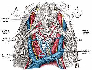
Neck anatomy
11/10/2009 04:02:00 م
this image shows the major vessels in the neck region and their relations to each other showing: 1. sternothyroid m. 2. inf. thyroid vein 3. middle thyroid vein 4. cricothyroid m. 5. sternothyroid art... More Details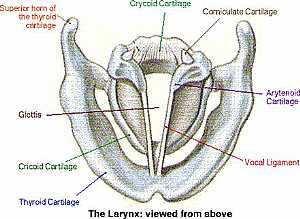
Larynx anatomy
12/10/2009 04:37:00 ص
this is superior view of the larynx showing the vocal cords showing: 1. vocal ligament 2. arytenoid cartilage 3. corniculate cartilage 4. crycoid cartilage 5. sup. horn of thyroid cartilage 6. glottis... More Details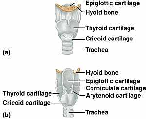
Larynx anatomy
12/10/2009 04:35:00 ص
the upper image is the larynx with thyroid cartilage on the lower image is after removing the anterior part of the thyroid cartilage showing: 1. epiglottic cartilage 2. hyoid bone 3. thyroid cartilage... More DetailsNeck anatomy
11/10/2009 04:02:00 م
this image shows the major vessels in the neck region and their relations to each other showing: 1. sternothyroid m. 2. inf. thyroid vein 3. middle thyroid vein 4. cricothyroid m. 5. sternothyroid art... More Details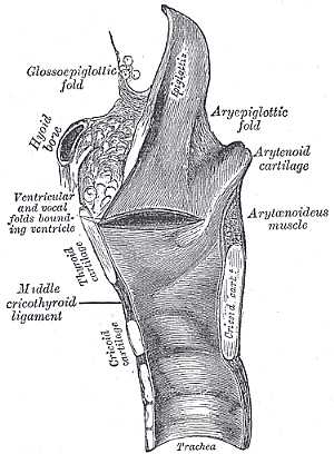
Larynx Anatomy
19/10/2009 03:06:37 م
In This Section you will find detailed different Photos and images about the anatomy of the Larynx including its surface , attachments , related structures , vocal cords and many more Items about the... More DetailsLarynx anatomy
12/10/2009 04:35:00 ص
the upper image is the larynx with thyroid cartilage on the lower image is after removing the anterior part of the thyroid cartilage showing: 1. epiglottic cartilage 2. hyoid bone 3. thyroid cartilage... More DetailsRelated Searches
loading...
loading...
Top Health & Medical Articles
New Articles
Most Read
آخر كلمات البحث
orthopaedic joint assessment centr dr mcmahon
, , , , , , ,anatomi ligamen panggul wanita
, ,abdomen sans preparation normale
, , , , , ,world conferences on urine therapy
, , , , , , , , , ,eDoctorOnline.com does not provide medical advice, diagnosis or treatment.
© Copyright 2001-2022 eDoctorOnline.com
© Copyright 2001-2022 eDoctorOnline.com