vertebra axis
axis vertebra, axis vertebrae, vertebra axis, odontoid process, axis vertebra, Axis c2, axis vertebrae, groove for vertebral artery, c2 axis, axis vertebra, atlas vertebra, atlas vertebra, atlas groove for vertebral artery, C2 AXIS, vertebra axis, odontoid process of axis, vertebra, atlas vertebrae, superior view of axis, vertebra C2,
The following are the result pages for the searched keyowrd: vertebra axis
the axis vertebra
10/10/2009 03:59:00 م
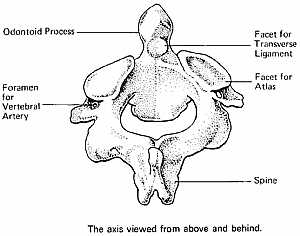
this is a detailed superior view of the second vertebra [C2] [the axis] showing: 1. odontoid process 2. facet for transverse ligament 3. facet for atlas 4. spine 5. foramen for vertebral artery... More Detailsthe axis vertebra
10/10/2009 03:59:00 م
this is a detailed superior view of the second vertebra [C2] [the axis] showing: 1. odontoid process 2. facet for transverse ligament 3. facet for atlas 4. spine 5. foramen for vertebral artery... More Details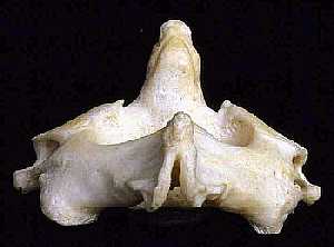
the Axis vertebra
10/10/2009 03:53:00 م
this is frontal view of the second vertebra in the vertebral column C2 : the second cervical vertebra [the axis]... More Details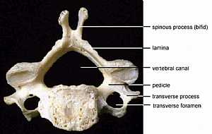
Vertebral Anatomy
19/10/2009 03:02:39 م
In This Section you will find detailed different Photos and images about the anatomy of the Vertebrae bones including their types , their surface , attachments related structures many more Items about... More Details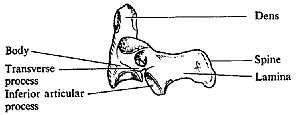
the axis vertebra
10/10/2009 03:58:00 م
this is a detailed lateral view of the axis vertebra [C2] showing: 1. dens 2. spine 3. lamina 4. body 5. transverse process 6. inf. auricular process... More Detailsthe Axis vertebra
10/10/2009 03:53:00 م
this is frontal view of the second vertebra in the vertebral column C2 : the second cervical vertebra [the axis]... More Detailsthe axis vertebra
10/10/2009 03:58:00 م
this is a detailed lateral view of the axis vertebra [C2] showing: 1. dens 2. spine 3. lamina 4. body 5. transverse process 6. inf. auricular process... More Details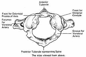
the first vertebra
10/10/2009 03:56:00 م
this is a detailed image for the first vertebra [ the atlas] in the vertebral column showing: 1. ant. tubercle 2. facet of occipital condyle 3. groove for vertebral artery 4. post. tubercle representi... More Detailsthe first vertebra
10/10/2009 03:56:00 م
this is a detailed image for the first vertebra [ the atlas] in the vertebral column showing: 1. ant. tubercle 2. facet of occipital condyle 3. groove for vertebral artery 4. post. tubercle representi... More Details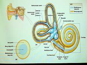
inner ear anatomy
12/10/2009 04:19:00 ص
this is a detailed image for the inner ear showing: 1. semicircular canals 2. semicircular ducts ( ant , post and middle) 3. vestibule 4. ampullae 5. maculae 6. endolymphatic sac 7. utricle 8. saccule... More Details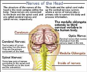
The nervous system nersves
16/10/2009 02:02:00 م
this image shows the brain stem with the nerves emerging from it showing: 1. cerebrum 2. medulla oblongata 3. optic nerve 4. cerebral nerves 5. spinal nerves... More DetailsRelated Searches
loading...
loading...
Top Health & Medical Articles
New Articles
Most Read
آخر كلمات البحث
orthopaedic joint assessment centr dr mcmahon
, , , , , , ,anatomi ligamen panggul wanita
, ,abdomen sans preparation normale
, , , , , ,world conferences on urine therapy
, , , , , , , , , ,eDoctorOnline.com does not provide medical advice, diagnosis or treatment.
© Copyright 2001-2022 eDoctorOnline.com
© Copyright 2001-2022 eDoctorOnline.com