vertebral column
spinal cord anatomy, spinal cord, spinal cord, spinal cord anatomy, cns, CNS, transverse foramen, cns anatomy, skeletal system, image, anatomy of the spine, spinal column anatomy, human skeletal system, CNS anatomy, spinal cord anatomy, human spinal cord, central nervous system anatomy, human spinal cord, anatomy of central nervous system, central nervous system anatomy,
The following are the result pages for the searched keyowrd: vertebral column
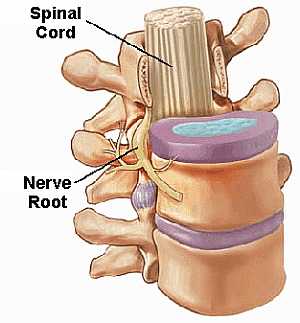
Anatomy of the Spine
05/03/2009 03:27:00 م
this image shows the spinal cord being protected inside the vertebral column showing: 1. spinal cord 2. nerve root 3. vertebral column... More DetailsAnatomy of the Spine
05/03/2009 03:27:00 م
this image shows the spinal cord being protected inside the vertebral column showing: 1. spinal cord 2. nerve root 3. vertebral column... More Details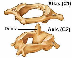
The Atlas and The Axis vertebrae
10/10/2009 03:50:00 م
this the first two vertebrae in the vertebral column the first (the atlas) and the second ( the axis) and shows how the two are put together and articulate together... More Details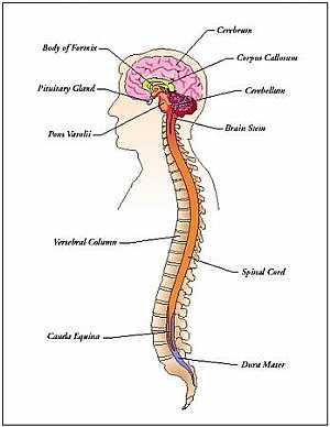
C.N.S anatomy
23/10/2009 04:01:00 م
this image shows the whole central nervous system in relation to each other showing: 1. cerebrum 2. corpus callosum 3. cerebellum 4. pituitary gland 5. brain stem 6. spinal cord 7. vertebral column 8... More Details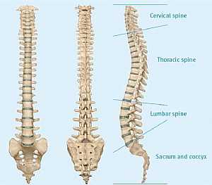
Spinal cord anatomy
23/10/2009 03:51:00 م
this shows the different segments of the vertebral column showing: 1. cervical 2. thoracic 3. lumbar 4. sacral 5. coccygeal... More Details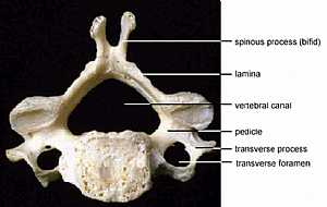
Vertebral Anatomy
19/10/2009 03:02:39 م
In This Section you will find detailed different Photos and images about the anatomy of the Vertebrae bones including their types , their surface , attachments related structures many more Items about... More DetailsSpinal cord anatomy
22/10/2009 01:52:10 م
In This Section you will find detailed different Photos and images about the anatomy of the Spinal cord including its surface , parts , related structures , Functions of the spinal; cord , Spinal nerv... More Details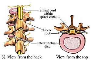
Spinal cord nerves anatomy
23/10/2009 03:20:00 م
this image shows the relation of the spinal cord to the vertebral column showing: 1. spinal cord 2. spinal canal 3. nerve root 4. intervertebral disc... More DetailsAnatomy of the vertebral column
10/10/2009 04:16:00 م
this is a typical cervical vertebra the typical cervical vertebra is from C3 to C6 showing: 1. spine (bifid) 2. lamina 3. pedicle 4. vertebral canal 5. transverse process 6. transverse foramen... More Details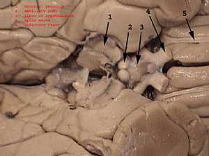
Hypothalamus anatomy
13/10/2009 01:03:00 ص
in this image the two cerebri are widened from each other to show the structure just beneath them ( the Hypothalamus) showing: 1. cerebral peduncle 2. mamillary body 3. floor of hypothalamus 4. optic ... More DetailsThe Atlas and The Axis vertebrae
10/10/2009 03:50:00 م
this the first two vertebrae in the vertebral column the first (the atlas) and the second ( the axis) and shows how the two are put together and articulate together... More Details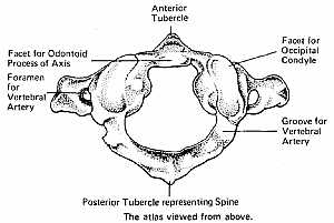
the first vertebra
10/10/2009 03:56:00 م
this is a detailed image for the first vertebra [ the atlas] in the vertebral column showing: 1. ant. tubercle 2. facet of occipital condyle 3. groove for vertebral artery 4. post. tubercle representi... More Details
C.N.S anatomy
23/10/2009 04:01:00 م
this image shows the whole central nervous system in relation to each other showing: 1. cerebrum 2. corpus callosum 3. cerebellum 4. pituitary gland 5. brain stem 6. spinal cord 7. vertebral column 8... More DetailsAnatomy of the vertebral column
10/10/2009 04:16:00 م
this is a typical cervical vertebra the typical cervical vertebra is from C3 to C6 showing: 1. spine (bifid) 2. lamina 3. pedicle 4. vertebral canal 5. transverse process 6. transverse foramen... More Details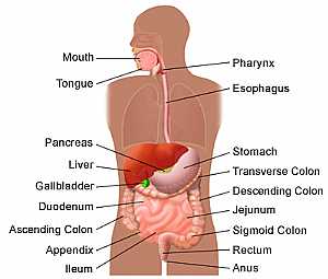
Systemic anatomy of the human body
11/11/2009 03:03:45 م
In This Section you will find detailed different Photos and images about the Different Systems of the human body including Nervous system , The circulatory system , Skeletal System and many more Item... More Details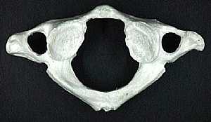
The Atlas vertebra
10/10/2009 03:47:00 م
this is the first vertebra in the vertebral column C1 : cervical number one [the Atlas]... More Detailsthe first vertebra
10/10/2009 03:56:00 م
this is a detailed image for the first vertebra [ the atlas] in the vertebral column showing: 1. ant. tubercle 2. facet of occipital condyle 3. groove for vertebral artery 4. post. tubercle representi... More Details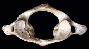
Anatomy of the first vertebra
10/10/2009 04:01:00 م
this is a superior view of the atlas vertebra [ c1 ]... More Details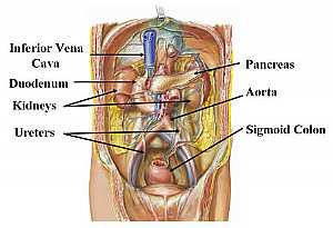
Abdomen anatomy
13/11/2009 05:58:00 ص
this image shows the abdomen from anterior view showing some of the abdominal organs in the abdominal region showing: 1. inferior vena cava 2. pancreas 3. sigmoid colon 4. ureters 5. kidneys 6. duode... More DetailsThe Atlas vertebra
10/10/2009 03:47:00 م
this is the first vertebra in the vertebral column C1 : cervical number one [the Atlas]... More Detailsorthopaedic joint assessment centr dr mcmahon
, , , , , , ,anatomi ligamen panggul wanita
, ,abdomen sans preparation normale
, , , , , ,world conferences on urine therapy
, , , , , , , , , ,© Copyright 2001-2022 eDoctorOnline.com