Solitary nucleus
septal area, stria terminalis, stria terminalis, septal area, cerebrum, baroreceptor reflex pathway, cerebrum, baroreceptor reflex pathway, cranial nerves, cranial nerves, otic ganglion, hippocampal formation, reticular formation, hippocampal formation, stria medullaris thalami, baroreceptor reflex, solitary nucleus, reticular formation, hypoglossal nerve, septal nuclei,
The following are the result pages for the searched keyowrd: Solitary nucleus
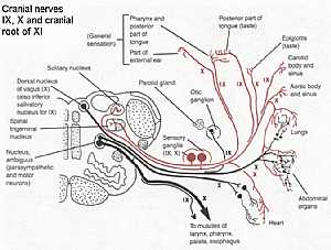
Cranial nerves IX,X,XI anatomy
28/10/2009 02:15:00 ص
this diagram shows the course of the cranial nerves( glossopharygeal IX,vagus X,and accessory XI ) from their origin to the their supplying organs showing: 1. solitary nucleus 2. spinal trigeminal nu... More Details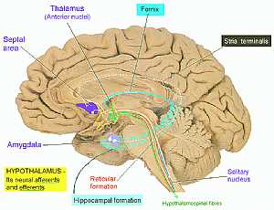
Cerebrum anatomy
16/10/2009 03:08:00 م
this is the medial wall of the cerebrum detailing the limbic system ( the system responsible for our behavior and thinking) showing: 1. septal area 2. thalamus 3. fornix 4. stria terminalis 5. solitar... More Details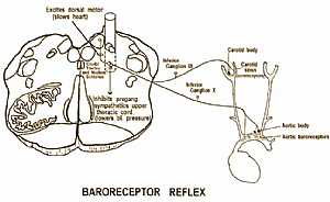
Baroreceptor reflex pathway
02/11/2009 02:20:00 م
this image shows the pathway of the baroreceptor reflex "the reflex that shares in the control of normal blood pressure" showing: 1. aortic baroreceptor 2. aortic body 3. carotid sinus 4. car... More DetailsBaroreceptor reflex pathway
02/11/2009 02:20:00 م
this image shows the pathway of the baroreceptor reflex "the reflex that shares in the control of normal blood pressure" showing: 1. aortic baroreceptor 2. aortic body 3. carotid sinus 4. car... More Details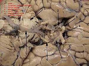
Occulomotor nerve anatomy
28/10/2009 02:10:00 ص
this image shows the oculomotor nerve at its origin in relation to the surrounding structures showing: 1. pons 2. internal carotid artery 3. optic nerve 4. optic chiasma 5. optic tract 6. occulomotor... More Details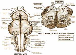
Hypoglossal nerve anatomy
03/11/2009 02:53:00 م
this image shows the hypoglossal nerve at its origin in relation to the surrounding structures showing: 1. hypoglossal nerve 2. cranial root of spinal accessory nerve 3. spinal root of spinal accesso... More Details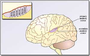
Cerebrum anatomy
22/10/2009 01:50:07 م
In This Section you will find detailed different Photos and images about the anatomy of the Cerebrum including its surface , parts , related structures , Functional areas of the brain , different cent... More Details
Cranial nerves anatomy
22/10/2009 01:51:53 م
In This Section you will find detailed different Photos and images about the anatomy of the Cranial Nerves including Their types , Fascial nerve anatomy , trigeminal nerve anatomy , vagus nerve anatom... More Details
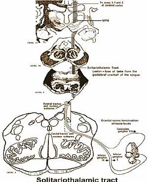
taste sensation pathway
02/11/2009 02:25:00 م
this image shows the solitariothalamic tract"the tract that connects the thalamus and the solitary nucleus" and responsible for the taste sensation showing: 1. taste buds in the tongue 2. cra... More Details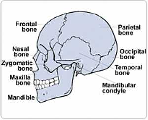
Skull - Latral Side
27/04/2006
Your skull is composed of many bones that enclose the brain and form your facial skeleton. showing: 1. frontal bone 2. parietal bone 3. occipital bone 4. temporal bone 5. mandibular bone 6. mandible... More DetailsCerebrum anatomy
16/10/2009 03:08:00 م
this is the medial wall of the cerebrum detailing the limbic system ( the system responsible for our behavior and thinking) showing: 1. septal area 2. thalamus 3. fornix 4. stria terminalis 5. solitar... More DetailsRelated Searches
loading...
loading...
Top Health & Medical Articles
New Articles
Most Read
آخر كلمات البحث
orthopaedic joint assessment centr dr mcmahon
, , , , , , ,anatomi ligamen panggul wanita
, ,abdomen sans preparation normale
, , , , , ,world conferences on urine therapy
, , , , , , , , , ,eDoctorOnline.com does not provide medical advice, diagnosis or treatment.
© Copyright 2001-2022 eDoctorOnline.com
© Copyright 2001-2022 eDoctorOnline.com