anatomy ear
nasal cavity, ear anatomy, ear anatomy, nose anatomy, NASAL ANATOMY, choana, anatomy of ear, sinus cavity, ear anatomy, pharyngeal tonsil, anatomy of nose, ear anatomy, anatomy of ear, sphenoid sinus, anatomy of the nose, nasal vestibule, inner ear anatomy, anatomy of ear, nasal turbinates, inner ear anatomy,
The following are the result pages for the searched keyowrd: anatomy ear
Ear anatomy
12/10/2009 04:21:00 ص
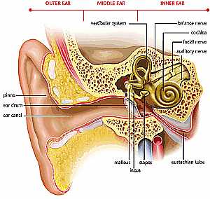
this image shows the different structures of the ear (external , middle and inner ear ) showing: 1. ear pinna 2. ear drum 3. ear canal 4. malleus 5. incus 6. stapes 7. eustacian tube 8. vestibular sy... More DetailsEar anatomy
12/10/2009 04:21:00 ص
this image shows the different structures of the ear (external , middle and inner ear ) showing: 1. ear pinna 2. ear drum 3. ear canal 4. malleus 5. incus 6. stapes 7. eustacian tube 8. vestibular sy... More Details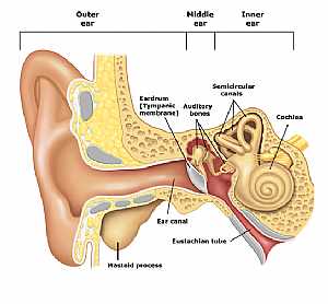
Ear anatomy
12/10/2009 04:42:00 ص
this shows the structure of the ear outer,middle and inner ear adjoined together showing: 1. mastoid process 2. ear canal 3. Eustachian tube 4. ear drum 5. auditory nerves 6. semicircular canals 7. co... More DetailsEar anatomy
12/10/2009 04:42:00 ص
this shows the structure of the ear outer,middle and inner ear adjoined together showing: 1. mastoid process 2. ear canal 3. Eustachian tube 4. ear drum 5. auditory nerves 6. semicircular canals 7. co... More Details
Ear anatomy
12/10/2009 04:40:00 ص
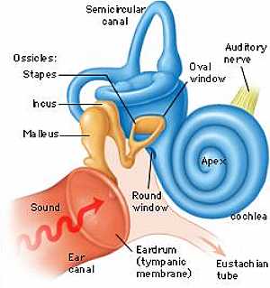
this is the middle and the inner ear together with their detailed structures showing: 1. ear canal 2. ear drum 3. Eustachian tube 4. malleus 5. incus 6. stapes 7. round window 8. oval window 9. audito... More Details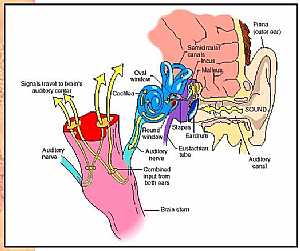
Ear anatomy
12/10/2009 04:19:00 ص
this image shows the pathway of the nerve impulses that come from the inner ear how to get to the brain showing: 1. ear drum 2. malleus bone 3. incus bone 4. stapes bone 5. Eustachian tube 6. semicirc... More Details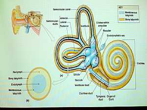
inner ear anatomy
12/10/2009 04:19:00 ص
this is a detailed image for the inner ear showing: 1. semicircular canals 2. semicircular ducts ( ant , post and middle) 3. vestibule 4. ampullae 5. maculae 6. endolymphatic sac 7. utricle 8. saccule... More DetailsEar anatomy
12/10/2009 04:19:00 ص
this image shows the pathway of the nerve impulses that come from the inner ear how to get to the brain showing: 1. ear drum 2. malleus bone 3. incus bone 4. stapes bone 5. Eustachian tube 6. semicirc... More Details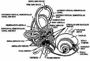
inner ear anatomy
12/10/2009 04:28:00 ص
this is a detailed image for the inner ear showing: 1. endolymph space 2. perilymph space 3. post. vertical semicircular canal and duct 4. membranous ampulla 5. horizontal semicircular canal and duct ... More Details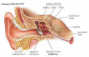
Ear anatomy
12/10/2009 04:22:00 ص
this image details the different structures of the ear specially the inner ear showing: 1. external auditory canal 2. tympanic membrane 3. tympanic cavity 4. malleus 5. incus 6. stapes 7. semicircular... More Details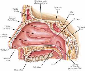
Nose anatomy
11/10/2009 03:55:00 م
this is a longitudinal section in the nasal cavity showing its medial wall showing: 1. frontal sinus 2. superior turbinate 3. middle turbinate 4. inferior turbinate 5. vestibule 6. hard palate 7. soft... More Detailsinner ear anatomy
12/10/2009 04:19:00 ص
this is a detailed image for the inner ear showing: 1. semicircular canals 2. semicircular ducts ( ant , post and middle) 3. vestibule 4. ampullae 5. maculae 6. endolymphatic sac 7. utricle 8. saccule... More DetailsRelated Searches
loading...
loading...
Top Health & Medical Articles
New Articles
Most Read
آخر كلمات البحث
orthopaedic joint assessment centr dr mcmahon
, , , , , , ,anatomi ligamen panggul wanita
, ,abdomen sans preparation normale
, , , , , ,world conferences on urine therapy
, , , , , , , , , ,eDoctorOnline.com does not provide medical advice, diagnosis or treatment.
© Copyright 2001-2022 eDoctorOnline.com
© Copyright 2001-2022 eDoctorOnline.com