anatomy of brain
brain anatomy, midbrain, forebrain, skull anatomy, hindbrain, corpus callosum, diencephalon, thalamus, forebrain midbrain hindbrain, anatomy of the brain, brain anatomy, thalamus anatomy, anatomy of the brain, brain anatomy, brain anatomy, image, brain anatomy, anatomy of brain, cranial nerves, midbrain anatomy,
The following are the result pages for the searched keyowrd: anatomy of brain
Brain anatomy
13/10/2009 12:33:00 ص
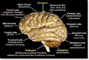
this is a detailed lateral view of the brain (cerebrum,cerebellum and brain stem) showing: 1. Brain stem 2. Cerebellum 3. Cerebrum 4. temporal lobe 5. frontal lobe 6. broca's area 7. frontal lobe ... More Details
Bones of Chest
16/07/2010 05:11:44 ص
In This Section you will find detailed different Photos and images about the anatomy of the Bones of the chest including its surface , parts , related structures and many more Items about the thoracic... More Details
Cerebrum anatomy
22/10/2009 01:50:07 م
In This Section you will find detailed different Photos and images about the anatomy of the Cerebrum including its surface , parts , related structures , Functional areas of the brain , different cent... More DetailsBrain anatomy
13/10/2009 12:33:00 ص
this is a detailed lateral view of the brain (cerebrum,cerebellum and brain stem) showing: 1. Brain stem 2. Cerebellum 3. Cerebrum 4. temporal lobe 5. frontal lobe 6. broca's area 7. frontal lobe ... More Details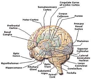
Brain anatomy
13/10/2009 12:50:00 ص
this is a detailed image of the cerebrum centers and its place on the cerebrum showing: 1. spinal cord 2. substantia nigra 3. optic t5ract 4. pituitary gland 5. hippocampus 6. hypothalamus 7. optic ne... More Details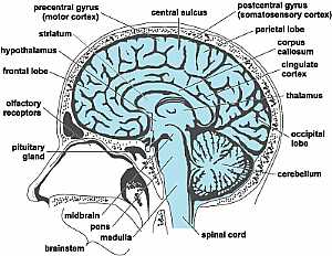
Head anatomy
16/10/2009 02:03:00 م
this is a longitudinal section in the head showing the brain and the brain stem showing: 1. pituitary gland 2. olfactory receptors 3. frontal lobe 4. hypothalamus 5. striatum 6. precentral gyrus 7. ce... More Details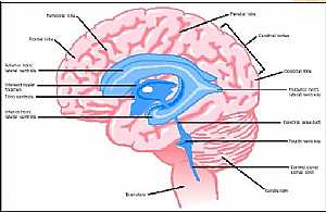
Brain anatomy
13/10/2009 12:36:00 ص
this is longitudinal cut section in the brain showing its parts the pink color is the nervous tissue and the blue color is what is called the ventricles ( spaces inside the brain filled with fluid ) s... More Details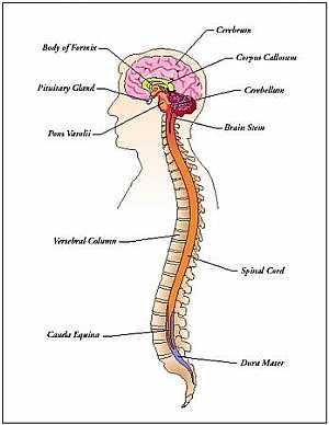
Nervous system and Special senses anatomy
12/10/2009 04:44:52 ص
In This Section you will find detailed different Sections about the different organs and structures in the Nervous System including The cerebrum , The cerebellums , The brain Stem , The ventricles of ... More Details
Atlas of Human Anatomy
27/04/2006
In This Section you will find detailed different sections about all different parts of the human body including head and neck , chest , abdomen , upper limbs , pelvis and lower limbs and many more Sec... More Details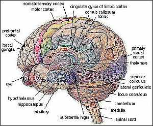
Brain anatomy
14/10/2009 04:18:00 ص
this is a detailed image for the lateral view of the cerebri and the cerebellum showing: 1. substania nigra 2. pituitary gland 3. hippocampus 4. hypothalamus 5. eye 6. basal ganglia 7. prefrontal cort... More Details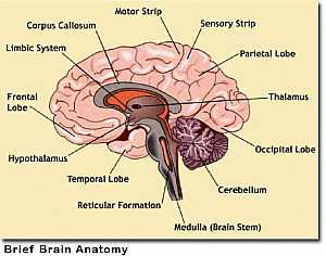
Cerebrum anatomy
13/10/2009 12:49:00 ص
this is a detailed image of the cerebrum and its parts showing: 1. temporal lobe 2. hypothalamus 3. frontal lobe 4. limbic system 5. corpus callosum 6. motor strip 7. sensory strip 8. parietal lobe 9.... More Details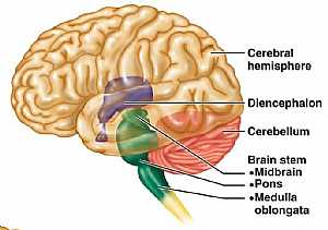
Brain anatomy
13/10/2009 12:39:00 ص
this is lateral view of the brain showing the cerebrum, cerebellum and their relation to the diencephalon (hypothalamus) and the brain stem (the root of the brain) showing: 1. cerebral hemisphere 2. d... More Details
Cranial nerves anatomy
22/10/2009 01:51:53 م
In This Section you will find detailed different Photos and images about the anatomy of the Cranial Nerves including Their types , Fascial nerve anatomy , trigeminal nerve anatomy , vagus nerve anatom... More Details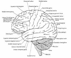
Brain anatomy
14/10/2009 04:34:00 ص
another lateral diagram for the sulci and gyri of the cerebrum showing: sulci: 1. lateral sulcus 2. inferior temporal sulcus 3. superior temporal sulcus 4. inferior frontal sulcus 5. superior frontal ... More Details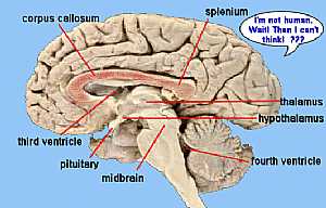
Brain anatomy
14/10/2009 04:26:00 ص
this is a longitudinal section in the brain showing the corpus callosum "the link between the two cerebri and shows the areas specialized in the process of "thinking" showing: 1. midbrain ... More DetailsBrain anatomy
13/10/2009 12:39:00 ص
this is lateral view of the brain showing the cerebrum, cerebellum and their relation to the diencephalon (hypothalamus) and the brain stem (the root of the brain) showing: 1. cerebral hemisphere 2. d... More Details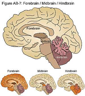
Brain anatomy
16/10/2009 02:05:00 م
this image differentiate between the forebrain and the midbrain and the hindbrain showing: 1. forebrain 2. midbrain 3. hindbrain... More Details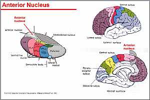
Thalamus anatomy
22/10/2009 02:21:00 م
this image shows the details of the thalamus and its nuclei showing: 1. anterior nuclei 2. ventral nuclei 3. geniculate body 4. lateral nuclei 5. medial nuclei 6. mediodorsal nucleus... More Details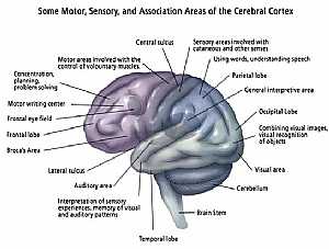
Brain anatomy
13/10/2009 12:46:00 ص
this image shows the main centers of the brain (every center has its own specific function) showing: 1. temporal lobe 2. auditory area 3. lateral sulcus 4. broca's area 5. frontal lobe 6. frontal ... More DetailsBrain anatomy
14/10/2009 04:18:00 ص
this is a detailed image for the lateral view of the cerebri and the cerebellum showing: 1. substania nigra 2. pituitary gland 3. hippocampus 4. hypothalamus 5. eye 6. basal ganglia 7. prefrontal cort... More Details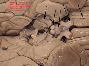
Hypothalamus anatomy
13/10/2009 01:03:00 ص
in this image the two cerebri are widened from each other to show the structure just beneath them ( the Hypothalamus) showing: 1. cerebral peduncle 2. mamillary body 3. floor of hypothalamus 4. optic ... More Details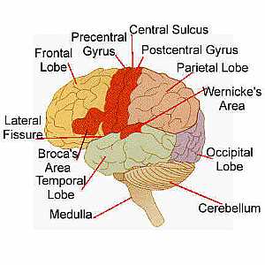
Brain anatomy
13/10/2009 01:04:00 ص
this is another image for the different areas of the cerebral hemisphere showing: 1. cerebellum 2. occipital lobe 3. wernicke's area 4. parietal lobe 5. postcentral gyrus 6. central sulcus 7. prec... More Details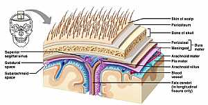
Brain anatomy
13/10/2009 01:14:00 ص
this image shows the coverings of the brain till the overlying skin showing: 1. skin of the scalp 2. periosteum 3. bone of the skull 4. Dura matter 5. arachnoid matter 6. pia matter 7. arachnoid villu... More Details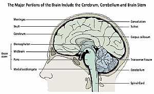
Head anatomy
13/10/2009 12:48:00 ص
this image shows the brain ,the brain stem and the spinal corn in their position in the head (skull) and neck (vertebral column) showing: 1. Brain stem 2. cerebrum 3. skull 4. meninges 5. Gyri 6. Sulc... More Details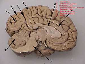
Cerebral hemisphere
13/10/2009 01:11:00 ص
this is a view of the medial wall of the the brain ( the cerebral hemisphere and the what under them) showing: 1. cerebellum 2. calcarine fissure 3. occipital lobe 4. parietoccipital fissure 5. pariet... More Details
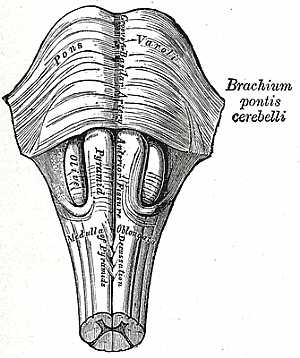
Brain stem anatomy
22/10/2009 01:50:57 م
In This Section you will find detailed different Photos and images about the anatomy of the Brain Stem including its surface , parts , related structures , midbrain anatomy , pons anatomy , medulla an... More Details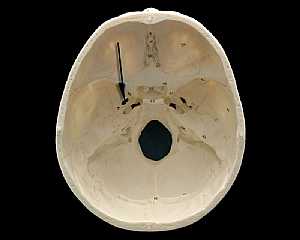
Skull Anatomy
19/10/2009 03:02:24 م
In This Section you will find detailed different Photos and images about the anatomy of the Skull bone including its surface , attachments related structures many more Items about the Skull anatomy... More Details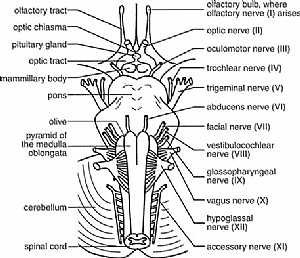
cranial nerves anatomy
14/10/2009 04:16:00 ص
this diagram shows the 12 cranial nerves and where they exactly exit from the brain showing: 1. all cranial nerves 2. olfactory bulb 3. optic chisma 4. pituitary gland 5. optic tract 6. mammillary bod... More Detailsorthopaedic joint assessment centr dr mcmahon
, , , , , , ,anatomi ligamen panggul wanita
, ,abdomen sans preparation normale
, , , , , ,world conferences on urine therapy
, , , , , , , , , ,© Copyright 2001-2022 eDoctorOnline.com