atlas of the human body
eye anatomy, eye diagram, human eye diagram, anatomy of eye, auditory pathway, diagram of the eye, human eye anatomy, body anatomy, anatomy eye, diagram of eye, ribs anatomy, anatomy of the eye, eye anatomy diagram, anatomy stomach, thoracic cage, ora serrata, abdomen anatomy, human eye, anatomy of the body, human anatomy diagram,
The following are the result pages for the searched keyowrd: atlas of the human body
Systemic anatomy of the human body
11/11/2009 03:03:45 م
In This Section you will find detailed different Photos and images about the Different Systems of the human body including Nervous system , The circulatory system , Skeletal System and many more Item... More Details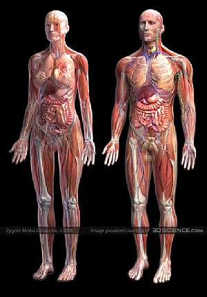
Muscles Anatomy (muscular system)
05/03/2009 02:10:00 م
this image shows the different muscles the forms and supports our human body and differs between male and female... More Details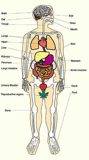
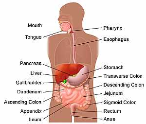
Anatomy of human body
12/11/2009 01:35:00 م
this image shows the human body from anterior view showing some organs of it showing: 1. brain 2. eye 3. nose 4. mouth 5. ear 6. throat 7. skin 8. lungs 9. heart 10. liver 11. kidneys 12. pancreas 13... More Details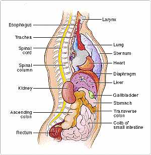
Body/Torso -- Side View (Body anatomy)
27/04/2006
Your torso consists of two parts — the chest and the abdomen. The chest contains your heart and lungs; your abdomen contains the digestive and urinary systems. Your chest and abdomen are separated by ... More Details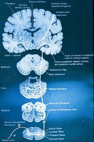
Pathways of C.N.S
22/10/2009 01:52:27 م
In This Section you will find detailed different Photos and images about the anatomy of the Pathways of the CNS including their types , spinothalamic track anatomy , ascending tracks anatomy , descend... More Details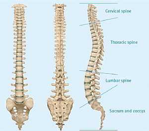
Spinal cord anatomy
22/10/2009 01:52:10 م
In This Section you will find detailed different Photos and images about the anatomy of the Spinal cord including its surface , parts , related structures , Functions of the spinal; cord , Spinal nerv... More Details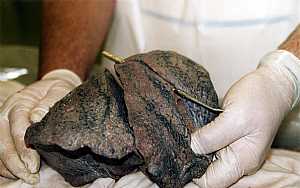
Lung anatomy
07/12/2009 06:31:00 ص
this image shows the anatomy of the right lung displaying its two main fissures the horizontal and the oblique one.they divide the right lung into three lobs "the upper lobe, the middle or the lat... More Details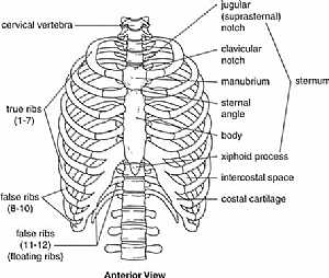
Thoracic cage anatomy
09/11/2009 03:42:00 م
this is an anterior view of the thoracic cage (the bones that forms the thorax) showing: 1. cervical vertebrae 2. jugular (suprasternal) notch 3. clavicular notch 4. manubrium 5. sternal angle 6. bod... More Details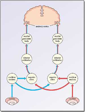
Auditory pathway
05/11/2009 03:41:00 ص
this image shows the pathway of the auditory system "the system responsible for our sense of hearing" (displays the pathway from both our left and right ears) showing: 1. cochlea "same si... More Details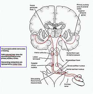
Auditory pathway
25/10/2009 03:19:00 م
this image shows the pathway of hearing system showing: 1. cochlear nerve 2. ventral cochlear nucleus 3. dorsal cochlear nucleus 4. olivocochlear fibers 5. sublentiform part of the internal capsule 6... More Details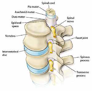
Spinal cord anatomy
16/10/2009 03:04:00 م
this image shows the relation of the spinal cord to its surrounding structures showing: 1. spinal cord 2. pia matter 3. arachnoid matter 4. dura matter 5. epidural space 6. vertebra 7. intervertebral ... More Details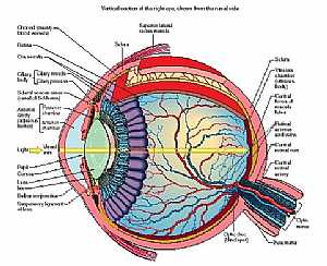
Eye anatomy
11/10/2009 04:00:00 م
this diagram details the different parts and structures of the human eye showing: 1. conjunctiva 2. ora serrata 3. cilliary body 4. aqueous 5. iris 6. ant. chamber 7. cornea 8. pupil 9. lens 10. post.... More Details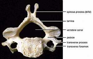
Anatomy of the vertebral column
10/10/2009 04:16:00 م
this is a typical cervical vertebra the typical cervical vertebra is from C3 to C6 showing: 1. spine (bifid) 2. lamina 3. pedicle 4. vertebral canal 5. transverse process 6. transverse foramen... More Details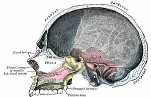
sagital section in the skull
09/10/2009 03:59:00 م
this is a longitudinal section in the skull viewing its lateral aspect (oral , nasal , frontal , parietal, occipital areas) showing: 1. frontal , prietal and occipital part of the skull 2. nasal bone ... More Details
Atlas of Human Anatomy
27/04/2006
In This Section you will find detailed different sections about all different parts of the human body including head and neck , chest , abdomen , upper limbs , pelvis and lower limbs and many more Sec... More Detailsorthopaedic joint assessment centr dr mcmahon
, , , , , , ,anatomi ligamen panggul wanita
, ,abdomen sans preparation normale
, , , , , ,world conferences on urine therapy
, , , , , , , , , ,© Copyright 2001-2022 eDoctorOnline.com