bones anatomy
femur, femur bone, skull anatomy, FIBULA, fibula, tibia fibula, fibula and tibia, tibia fibula, femur, tibia and fibula, FEMUR ANATOMY, metacarpal, fibula tibia, tibia bone, image, tibia and fibula, pelvic bone anatomy, femur bone anatomy, shoulder anatomy, metacarpal bones,
The following are the result pages for the searched keyowrd: bones anatomy
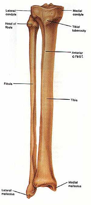
Fibula and Tibia bones anatomy
04/12/2009 07:43:00 ص
this image shows the anatomy of the fibula and tibia bones (the bones of the leg) in relation to each other ,displaying the different features and parts of them ,fibula (on the left) and the tibia (on... More Details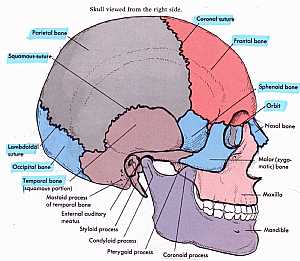
Skull Anatomy
10/10/2009 04:28:00 م
this shows the names of the bones that forms the side of the skull showing: 1. coronal suture 2. frontal bone 3. sphenoid bone 4. orbital bone 5. nasal bone 6. zygomatic bone 7. maxillary bone 8. mand... More Details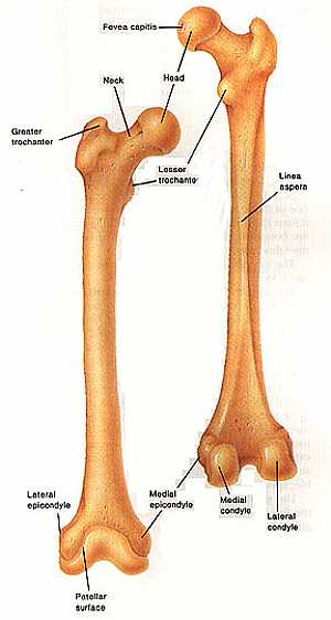
Femur bone anatomy
04/12/2009 07:30:00 ص
this image shows the anatomy of the femur bone from anterior and posterior view displaying its different features and parts showing: 1. fovea capitis 2. head of the femur 3. neck of the femur 4. less... More Details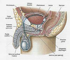
Pelvis Anatomy
06/11/2009 01:06:01 م
In This Section you will find detailed different Sections about the different organs and structures in the region of the Pelvis including Male pelvis anatomy , Female pelvis anatomy , pelvic girdle an... More Details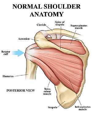
Shoulder joint anatomy
18/12/2009 07:20:00 ص
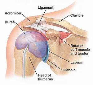
this image shows the anatomy of the shoulder joint from posterior view displaying the bones, tendons and muscles of the joint in relation to each other. showing: 1. Supraspinatus muscle 2. Spine of t... More DetailsLeg bones
15/07/2010 03:22:04 م
In This Section you will find detailed different Photos and images about the anatomy of the Leg bones including its surface , parts , related structures , attachments and many more Items about the Leg... More Details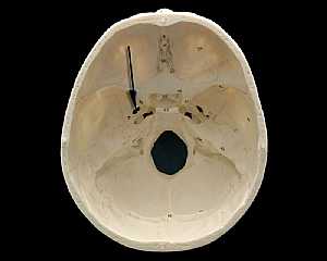
Skull Anatomy
19/10/2009 03:02:24 م
In This Section you will find detailed different Photos and images about the anatomy of the Skull bone including its surface , attachments related structures many more Items about the Skull anatomy... More Details
Shoulder Anatomy
14/07/2010 11:39:39 ص
In This Section you will find detailed different Photos and images about the anatomy of the Shoulder including its surface , parts , related structures , Muscles of the shoulder , Brachial plexus , te... More Details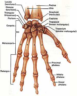
Hand anatomy
04/12/2009 06:23:00 ص
this image shows the anatomy of the hand displaying the bones forming the hand (carpal , metacarpal , phalanges bones) showing: 1. radius bone 2. ulna bone 3. scaphoid bone 4. capitate bone 5. trapez... More Details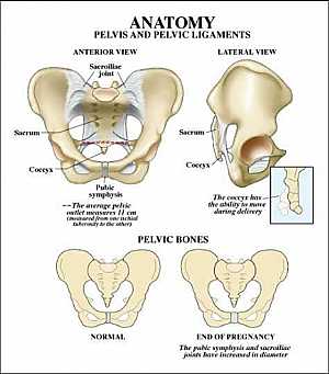
Pelvic bone anatomy
13/11/2009 06:32:00 ص
this image shows the pelvic bone that supports the pelvic region from anterior and lateral view showing: 1. sacroiliac joint 2. sacrum 3. coccyx 4. symphysis pubis 5. hib bone... More Details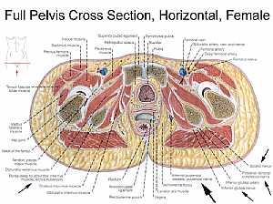
Female pelvic anatomy
13/11/2009 07:31:00 ص
this image is a horizontal section in a female pelvis shows the different pelvic organs and structures in relation to each other showing: 1. iliacus muscle 2. sartorius muscle 3. rectus femoris muscl... More DetailsRelated Searches
loading...
loading...
Top Health & Medical Articles
New Articles
Most Read
آخر كلمات البحث
orthopaedic joint assessment centr dr mcmahon
, , , , , , ,anatomi ligamen panggul wanita
, ,abdomen sans preparation normale
, , , , , ,world conferences on urine therapy
, , , , , , , , , ,eDoctorOnline.com does not provide medical advice, diagnosis or treatment.
© Copyright 2001-2022 eDoctorOnline.com
© Copyright 2001-2022 eDoctorOnline.com