brain anatomy
visual pathway, brain anatomy, midbrain, optic chiasm, forebrain, third ventricle, hindbrain, visual cortex, corpus callosum, diencephalon, thalamus, forebrain midbrain hindbrain, visual pathways, anatomy of the brain, brain anatomy, transverse fissure, thalamus anatomy, anatomy of the brain, cranial nerves, brain anatomy,
The following are the result pages for the searched keyowrd: brain anatomy
Brain anatomy
13/10/2009 12:33:00 ص
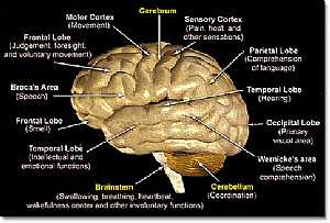
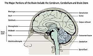
this is a detailed lateral view of the brain (cerebrum,cerebellum and brain stem) showing: 1. Brain stem 2. Cerebellum 3. Cerebrum 4. temporal lobe 5. frontal lobe 6. broca's area 7. frontal lobe ... More Details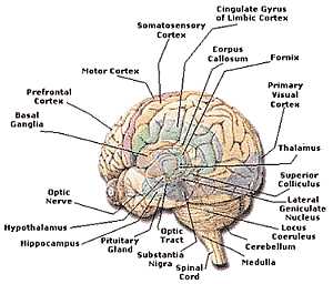
Brain anatomy
13/10/2009 12:50:00 ص
this is a detailed image of the cerebrum centers and its place on the cerebrum showing: 1. spinal cord 2. substantia nigra 3. optic t5ract 4. pituitary gland 5. hippocampus 6. hypothalamus 7. optic ne... More Details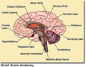
Cerebrum anatomy
13/10/2009 12:49:00 ص
this is a detailed image of the cerebrum and its parts showing: 1. temporal lobe 2. hypothalamus 3. frontal lobe 4. limbic system 5. corpus callosum 6. motor strip 7. sensory strip 8. parietal lobe 9.... More DetailsBrain anatomy
13/10/2009 12:33:00 ص
this is a detailed lateral view of the brain (cerebrum,cerebellum and brain stem) showing: 1. Brain stem 2. Cerebellum 3. Cerebrum 4. temporal lobe 5. frontal lobe 6. broca's area 7. frontal lobe ... More Details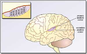
Cerebrum anatomy
22/10/2009 01:50:07 م
In This Section you will find detailed different Photos and images about the anatomy of the Cerebrum including its surface , parts , related structures , Functional areas of the brain , different cent... More Details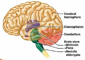
Brain anatomy
13/10/2009 12:39:00 ص
this is lateral view of the brain showing the cerebrum, cerebellum and their relation to the diencephalon (hypothalamus) and the brain stem (the root of the brain) showing: 1. cerebral hemisphere 2. d... More Details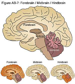
Brain anatomy
16/10/2009 02:05:00 م
this image differentiate between the forebrain and the midbrain and the hindbrain showing: 1. forebrain 2. midbrain 3. hindbrain... More Details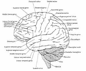
Brain anatomy
14/10/2009 04:34:00 ص
another lateral diagram for the sulci and gyri of the cerebrum showing: sulci: 1. lateral sulcus 2. inferior temporal sulcus 3. superior temporal sulcus 4. inferior frontal sulcus 5. superior frontal ... More Details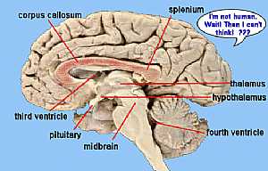
Brain anatomy
14/10/2009 04:26:00 ص
this is a longitudinal section in the brain showing the corpus callosum "the link between the two cerebri and shows the areas specialized in the process of "thinking" showing: 1. midbrain ... More Details
Bones of Chest
16/07/2010 05:11:44 ص
In This Section you will find detailed different Photos and images about the anatomy of the Bones of the chest including its surface , parts , related structures and many more Items about the thoracic... More DetailsBrain anatomy
13/10/2009 12:39:00 ص
this is lateral view of the brain showing the cerebrum, cerebellum and their relation to the diencephalon (hypothalamus) and the brain stem (the root of the brain) showing: 1. cerebral hemisphere 2. d... More DetailsBrain anatomy
16/10/2009 02:05:00 م
this image differentiate between the forebrain and the midbrain and the hindbrain showing: 1. forebrain 2. midbrain 3. hindbrain... More Details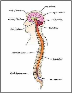
Nervous system and Special senses anatomy
12/10/2009 04:44:52 ص
In This Section you will find detailed different Sections about the different organs and structures in the Nervous System including The cerebrum , The cerebellums , The brain Stem , The ventricles of ... More Details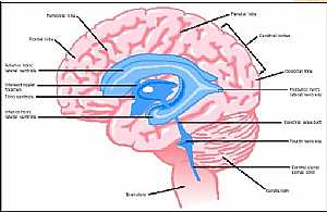
Brain anatomy
13/10/2009 12:36:00 ص
this is longitudinal cut section in the brain showing its parts the pink color is the nervous tissue and the blue color is what is called the ventricles ( spaces inside the brain filled with fluid ) s... More DetailsBrain anatomy
14/10/2009 04:34:00 ص
another lateral diagram for the sulci and gyri of the cerebrum showing: sulci: 1. lateral sulcus 2. inferior temporal sulcus 3. superior temporal sulcus 4. inferior frontal sulcus 5. superior frontal ... More Details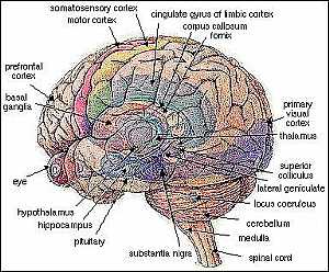
Brain anatomy
14/10/2009 04:18:00 ص
this is a detailed image for the lateral view of the cerebri and the cerebellum showing: 1. substania nigra 2. pituitary gland 3. hippocampus 4. hypothalamus 5. eye 6. basal ganglia 7. prefrontal cort... More Details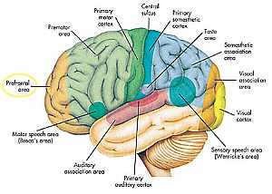
Cerebrum anatomy
12/10/2009 04:55:00 ص
God has said in the Quran about one of the evil unbelievers who forbade the Prophet Muhammad from praying at the Kaaba "No! If he does not stop, We will take him by the naseyah (front of the hea... More DetailsBrain anatomy
14/10/2009 04:18:00 ص
this is a detailed image for the lateral view of the cerebri and the cerebellum showing: 1. substania nigra 2. pituitary gland 3. hippocampus 4. hypothalamus 5. eye 6. basal ganglia 7. prefrontal cort... More Details
Head anatomy
13/10/2009 12:48:00 ص
this image shows the brain ,the brain stem and the spinal corn in their position in the head (skull) and neck (vertebral column) showing: 1. Brain stem 2. cerebrum 3. skull 4. meninges 5. Gyri 6. Sulc... More Details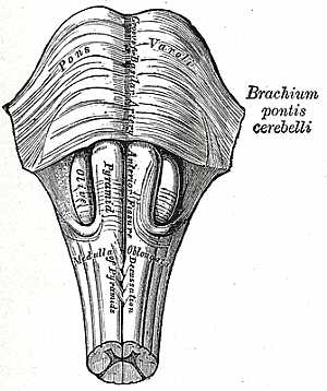
Brain stem anatomy
22/10/2009 01:50:57 م
In This Section you will find detailed different Photos and images about the anatomy of the Brain Stem including its surface , parts , related structures , midbrain anatomy , pons anatomy , medulla an... More Details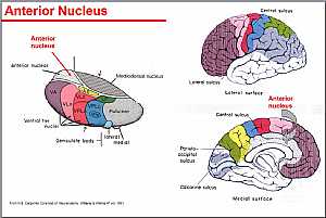
Thalamus anatomy
22/10/2009 02:21:00 م
this image shows the details of the thalamus and its nuclei showing: 1. anterior nuclei 2. ventral nuclei 3. geniculate body 4. lateral nuclei 5. medial nuclei 6. mediodorsal nucleus... More Details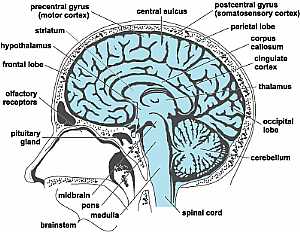
Head anatomy
16/10/2009 02:03:00 م
this is a longitudinal section in the head showing the brain and the brain stem showing: 1. pituitary gland 2. olfactory receptors 3. frontal lobe 4. hypothalamus 5. striatum 6. precentral gyrus 7. ce... More DetailsBrain anatomy
13/10/2009 12:36:00 ص
this is longitudinal cut section in the brain showing its parts the pink color is the nervous tissue and the blue color is what is called the ventricles ( spaces inside the brain filled with fluid ) s... More Details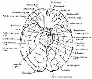
Brain anatomy
14/10/2009 04:43:00 ص
an inferior view of the brain with details on the different parts and areas on it showing: 1. rectus gyrus 2. olfactory sulcus 3. orbital gyri 4. optic nerve 5. optic chiasma 6. optic tract 7. infundi... More Details
Atlas of Human Anatomy
27/04/2006
In This Section you will find detailed different sections about all different parts of the human body including head and neck , chest , abdomen , upper limbs , pelvis and lower limbs and many more Sec... More Details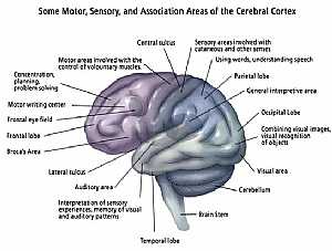
Brain anatomy
13/10/2009 12:46:00 ص
this image shows the main centers of the brain (every center has its own specific function) showing: 1. temporal lobe 2. auditory area 3. lateral sulcus 4. broca's area 5. frontal lobe 6. frontal ... More Details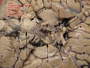
Brain anatomy
13/10/2009 01:08:00 ص
this image shows the back of the brain stem and the structures under the cerebri with the major vessels on them showing: 1. pons 2. internal carotid artery 3. optic nerve 4. optic chiasma 5. optic tra... More Details
Upper Limb Anatomy
06/11/2009 01:04:46 م
In This Section you will find detailed different Sections about the different organs and structures in the region of the Upper limbs including The shoulder , the arms , the forearms , the hand anatomy... More Details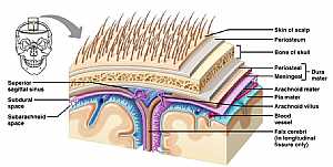
Brain anatomy
13/10/2009 01:14:00 ص
this image shows the coverings of the brain till the overlying skin showing: 1. skin of the scalp 2. periosteum 3. bone of the skull 4. Dura matter 5. arachnoid matter 6. pia matter 7. arachnoid villu... More Details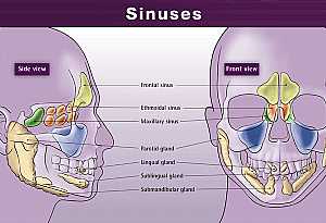
Head anatomy
11/10/2009 04:19:00 م
this shows the air sinuses of the skull ( these are spaces inside the skull bones filled with air) showing: 1. frontal sinus (yellow area) 2. ethmoidal sinus (orange are) 3. maxillary sinus (blue area... More Details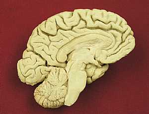
Brain anatomy
23/10/2009 03:03:00 م
this the left half of the brain (showing the medial wall of the left half)... More DetailsHead anatomy
16/10/2009 02:03:00 م
this is a longitudinal section in the head showing the brain and the brain stem showing: 1. pituitary gland 2. olfactory receptors 3. frontal lobe 4. hypothalamus 5. striatum 6. precentral gyrus 7. ce... More Details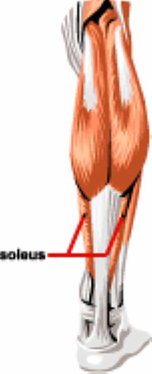
Leg muscles anatomy
15/07/2010 03:21:41 م
In This Section you will find detailed different Photos and images about the anatomy of the Leg muscles including its surface , parts , related structures , different muscles of the lower limbs and ma... More Details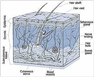
Brain anatomy
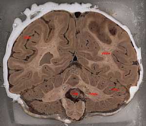
14/10/2009 04:21:00 ص
this is cut section in the brain vertically "in the image is the ant. part of the section" this image shows what is called the grey matter of the nervous system ( the cell bodies) and the whit... More DetailsBrain anatomy
23/10/2009 03:03:00 م
this the left half of the brain (showing the medial wall of the left half)... More DetailsBrain anatomy
14/10/2009 04:43:00 ص
an inferior view of the brain with details on the different parts and areas on it showing: 1. rectus gyrus 2. olfactory sulcus 3. orbital gyri 4. optic nerve 5. optic chiasma 6. optic tract 7. infundi... More Details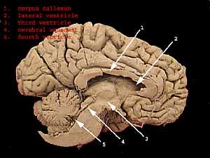
Brain ventricles
13/10/2009 01:20:00 ص
this section that shows the medial wall of the cerebral hemisphere shows the main four ventricles of the brain showing: 1. corpus callosum 2. lateral ventricle 3. cerebral aquiduct 4. fourth ventricle... More Details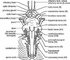
cranial nerves anatomy
14/10/2009 04:16:00 ص
this diagram shows the 12 cranial nerves and where they exactly exit from the brain showing: 1. all cranial nerves 2. olfactory bulb 3. optic chisma 4. pituitary gland 5. optic tract 6. mammillary bod... More Details
Cranial nerves anatomy
22/10/2009 01:51:53 م
In This Section you will find detailed different Photos and images about the anatomy of the Cranial Nerves including Their types , Fascial nerve anatomy , trigeminal nerve anatomy , vagus nerve anatom... More DetailsBrain anatomy
13/10/2009 12:46:00 ص
this image shows the main centers of the brain (every center has its own specific function) showing: 1. temporal lobe 2. auditory area 3. lateral sulcus 4. broca's area 5. frontal lobe 6. frontal ... More DetailsCerebrum anatomy
13/10/2009 12:49:00 ص
this is a detailed image of the cerebrum and its parts showing: 1. temporal lobe 2. hypothalamus 3. frontal lobe 4. limbic system 5. corpus callosum 6. motor strip 7. sensory strip 8. parietal lobe 9.... More Details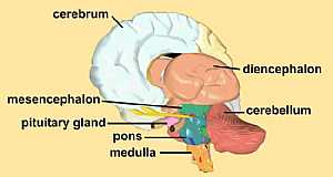
Brain anatomy
23/10/2009 03:05:00 م
this image shows the whole brain detailing its parts showing: 1. cerebrum 2. diencephalon 3. cerebellum 4. mesencephalon 5. piyuitary gland 6. pons 7. medulla... More Details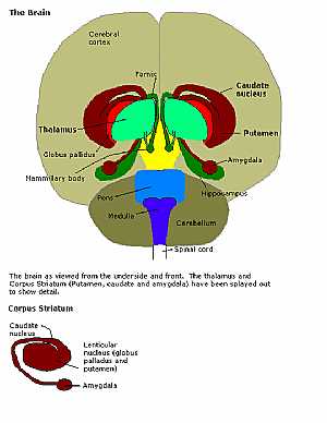
brain stem anatomy
23/10/2009 03:14:00 م
this is a anterior view of the whole brain showing the relation of the different parts of the brain stem to the rest of the brain showing: 1. cerebrum 2. fornix 3. thalamus 4. globus pallidus 5. mamm... More DetailsCranial nerves anatomy
05/11/2009 04:03:00 ص
this image shows the all cranial nerves and displaying their effector organs showing: 1. Olfactory nerve I 2. Optic nerve II 3. Occulomotor nerve III 4. Trochlear nerve IV 5. Trigeminal nerve V 6. Ab... More Details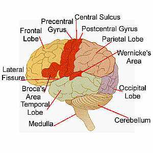
Brain anatomy
13/10/2009 01:04:00 ص
this is another image for the different areas of the cerebral hemisphere showing: 1. cerebellum 2. occipital lobe 3. wernicke's area 4. parietal lobe 5. postcentral gyrus 6. central sulcus 7. prec... More Details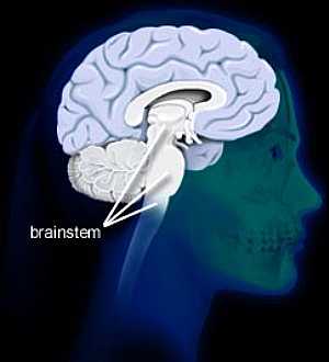
Brain stem anatomy
16/10/2009 02:00:00 م
this image shows the relation between the brain stem and the cerebri... More DetailsBrain anatomy
14/10/2009 04:26:00 ص
this is a longitudinal section in the brain showing the corpus callosum "the link between the two cerebri and shows the areas specialized in the process of "thinking" showing: 1. midbrain ... More Details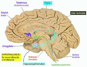
Cerebrum anatomy
16/10/2009 03:08:00 م
this is the medial wall of the cerebrum detailing the limbic system ( the system responsible for our behavior and thinking) showing: 1. septal area 2. thalamus 3. fornix 4. stria terminalis 5. solitar... More Details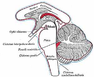
Ventricles of The C.N.S
22/10/2009 01:51:39 م
In This Section you will find detailed different Photos and images about the anatomy of the Ventricles of the brain including Their surfaces , parts , related structures and many more Items about the ... More DetailsBrain anatomy
13/10/2009 01:14:00 ص
this image shows the coverings of the brain till the overlying skin showing: 1. skin of the scalp 2. periosteum 3. bone of the skull 4. Dura matter 5. arachnoid matter 6. pia matter 7. arachnoid villu... More Details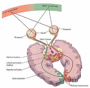
Visual pathway
03/11/2009 03:29:00 م
this image shows the visual pathway that carry sensation form the eye to the cerebral cortex showing: 1. temporal and nasal retina 2. optic nerve 3. optic chiasm 4. optic tract 5. pulvinar nucleus 6.... More Detailsorthopaedic joint assessment centr dr mcmahon
, , , , , , ,anatomi ligamen panggul wanita
, ,abdomen sans preparation normale
, , , , , ,world conferences on urine therapy
, , , , , , , , , ,© Copyright 2001-2022 eDoctorOnline.com