frontal lobe anatomy
brain anatomy, anatomy of the brain, brain anatomy, sylvian fissure, anatomy of brain, lateral fissure, PRECENTRAL GYRUS, central sulcus, anatomy, anatomy of brain, detailed brain anatomy, anatomy of the brain, the brain anatomy, detailed brain anatomy, human brain anatomy, brain fissures, brain anatomy, lateral sulcus, sensory strip, postcentral gyrus,
The following are the result pages for the searched keyowrd: frontal lobe anatomy
Brain anatomy
13/10/2009 12:33:00 ص
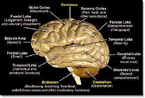
this is a detailed lateral view of the brain (cerebrum,cerebellum and brain stem) showing: 1. Brain stem 2. Cerebellum 3. Cerebrum 4. temporal lobe 5. frontal lobe 6. broca's area 7. frontal lobe ... More Details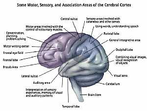
Brain anatomy
13/10/2009 12:46:00 ص
this image shows the main centers of the brain (every center has its own specific function) showing: 1. temporal lobe 2. auditory area 3. lateral sulcus 4. broca's area 5. frontal lobe 6. frontal ... More Details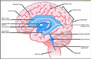
Brain anatomy
13/10/2009 12:36:00 ص
this is longitudinal cut section in the brain showing its parts the pink color is the nervous tissue and the blue color is what is called the ventricles ( spaces inside the brain filled with fluid ) s... More Details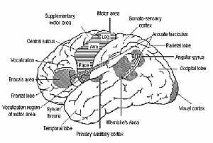
Cerebral hemispheres
13/10/2009 01:06:00 ص
this image shows what is called the sulci and gyri of the brain sulcus: is a groove in the brain tissue gyrus :is an elevation in the brain tissue note that the motor area represents the body upside ... More Details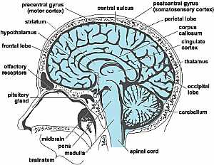
Head anatomy
16/10/2009 02:03:00 م
this is a longitudinal section in the head showing the brain and the brain stem showing: 1. pituitary gland 2. olfactory receptors 3. frontal lobe 4. hypothalamus 5. striatum 6. precentral gyrus 7. ce... More Details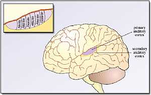
Cerebrum anatomy
22/10/2009 01:50:07 م
In This Section you will find detailed different Photos and images about the anatomy of the Cerebrum including its surface , parts , related structures , Functional areas of the brain , different cent... More DetailsBrain anatomy
13/10/2009 12:33:00 ص
this is a detailed lateral view of the brain (cerebrum,cerebellum and brain stem) showing: 1. Brain stem 2. Cerebellum 3. Cerebrum 4. temporal lobe 5. frontal lobe 6. broca's area 7. frontal lobe ... More Details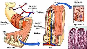
Small intestine anatomy
10/12/2009 09:14:00 ص
this image shows the anatomy of the wall of the small intestine showing what is called the villi of the small intestine (invagination of the wall of the small intestine into the lumen of the intestine... More Details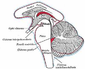
Ventricles of The C.N.S
22/10/2009 01:51:39 م
In This Section you will find detailed different Photos and images about the anatomy of the Ventricles of the brain including Their surfaces , parts , related structures and many more Items about the ... More Details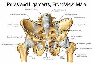
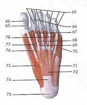
Lower limb anatomy
01/12/2009 06:52:00 ص
this image shows the anatomy of the foot from inferior view displaying mainly the muscles and tendons of the foot in relation to each other showing: 65. cruciform part of fibrous tendon sheaths 66. a... More DetailsRelated Searches
loading...
loading...
Top Health & Medical Articles
New Articles
Most Read
آخر كلمات البحث
orthopaedic joint assessment centr dr mcmahon
, , , , , , ,anatomi ligamen panggul wanita
, ,abdomen sans preparation normale
, , , , , ,world conferences on urine therapy
, , , , , , , , , ,eDoctorOnline.com does not provide medical advice, diagnosis or treatment.
© Copyright 2001-2022 eDoctorOnline.com
© Copyright 2001-2022 eDoctorOnline.com