larynx anatomy
larynx, mouth anatomy, larynx anatomy, stomach anatomy, larynx anatomy, oral cavity, stomach, pharynx anatomy, skull anatomy, cuneiform cartilage, larynx, cuneiform cartilage, larynx anatomy, larynx, Pharynx, larynx, anatomy of the mouth, anatomy of mouth, oral cavity anatomy, larynx anatomy,
The following are the result pages for the searched keyowrd: larynx anatomy
Larynx Anatomy
19/10/2009 03:06:37 م
In This Section you will find detailed different Photos and images about the anatomy of the Larynx including its surface , attachments , related structures , vocal cords and many more Items about the... More Details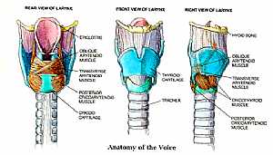
Larynx anatomy
12/10/2009 04:27:00 ص
this is a detailed image of the larynx from ant. and side view showing: 1. epiglottis 2. obique arytenoid m. 3. trans. and oblique arytenoid m. 4. post. cricoarytenoid m. 5. crcoid cartilage 6. thyroi... More DetailsLarynx anatomy
12/10/2009 04:27:00 ص
this is a detailed image of the larynx from ant. and side view showing: 1. epiglottis 2. obique arytenoid m. 3. trans. and oblique arytenoid m. 4. post. cricoarytenoid m. 5. crcoid cartilage 6. thyroi... More Details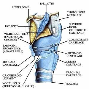
Larynx anatomy
12/10/2009 04:36:00 ص
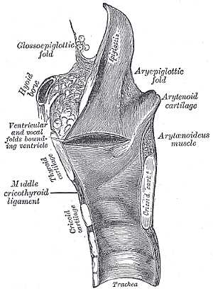
this is a longitudinal section in the trachea with side view detailing the structure of the larynx showing: 1. epiglottis 2. hyoid bone 3. fat body 4. vestibular fold(false vocal cord) 5. laryngeal pr... More Details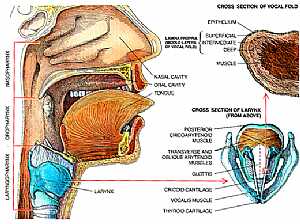
Pharynx and Larynx anatomy
12/10/2009 04:26:00 ص
this is a cut section in the head and the neck showing the laryngopharynx and the larynx on the right is a bigger image of the larynx with its detailed structure showing: 1. post. cricoarytenoid m. 2.... More Details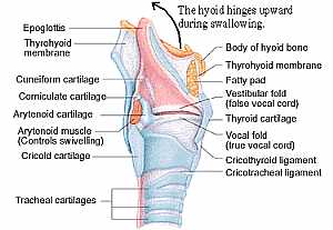
Anatomy of the larynx
11/10/2009 03:44:00 م
this is the structure of the larynx ( the throat area ) showing: 1. epiglottis 2. thyroid membrane 3. cuneiform cartilage 4. corniculate cartilage 5. arytenoid cartilage 6. arytenoid m. 7. cricoid car... More Details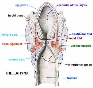
Larynx anatomy
12/10/2009 04:31:00 ص
this image shows the different structures of the larynx showing: 1. epiglottis 2. hyoid bone 3. thyroid cartilage 4. vocal ligament 5. crcoid cartilage 6. vestibule of the larynx 7. vestibular fold 8.... More DetailsLarynx anatomy
12/10/2009 04:31:00 ص
this image shows the different structures of the larynx showing: 1. epiglottis 2. hyoid bone 3. thyroid cartilage 4. vocal ligament 5. crcoid cartilage 6. vestibule of the larynx 7. vestibular fold 8.... More DetailsPharynx and Larynx anatomy
12/10/2009 04:26:00 ص
this is a cut section in the head and the neck showing the laryngopharynx and the larynx on the right is a bigger image of the larynx with its detailed structure showing: 1. post. cricoarytenoid m. 2.... More DetailsAnatomy of the larynx
11/10/2009 03:44:00 م
this is the structure of the larynx ( the throat area ) showing: 1. epiglottis 2. thyroid membrane 3. cuneiform cartilage 4. corniculate cartilage 5. arytenoid cartilage 6. arytenoid m. 7. cricoid car... More Details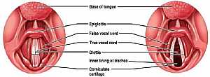
Larynx anatomy
12/10/2009 04:38:00 ص
this is the larynx from above with the vocal cords opened (right image) and closed (left image) showing: 1. base of the tongue 2. epiglottis 3. false vocal cord 4. true vocal cord 5. glottis 6. inner ... More Details
Larynx anatomy
12/10/2009 04:36:00 ص
this is a longitudinal section in the trachea with side view detailing the structure of the larynx showing: 1. epiglottis 2. hyoid bone 3. fat body 4. vestibular fold(false vocal cord) 5. laryngeal pr... More Details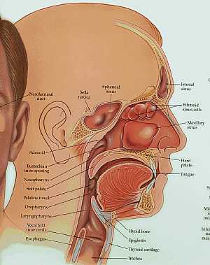
anatomy of the head and neck
09/10/2009 03:44:00 م
this image shows longitudinal section in the head and neck with their details (the frontal,nasal,oral and neck regions) showing: 1. nasolacrimal duct 2. sella turcica 3. sphenoid sinus 4. frontal sinu... More Details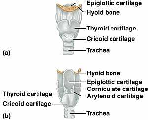
Larynx anatomy
12/10/2009 04:35:00 ص
the upper image is the larynx with thyroid cartilage on the lower image is after removing the anterior part of the thyroid cartilage showing: 1. epiglottic cartilage 2. hyoid bone 3. thyroid cartilage... More Details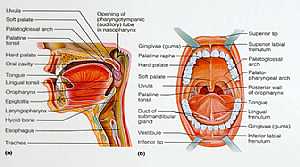
Oral Cavity
11/10/2009 04:09:00 م
the image on the right: is a detailed image of the structures of the mouth the image on the left: cut section in the head showing the structures of the mouth and oropharynx ( just after the mouth area... More Details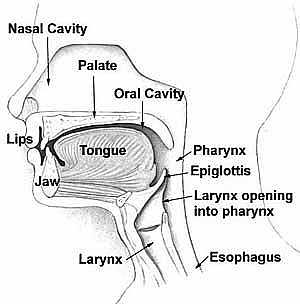
Head anatomy
11/10/2009 03:59:00 م
medial view of the nasal cavity, the mouth and the oropharynx part ( that,s the region just behind the tongue) showing: 1. nasal cavity 2. palate 3. oral cavity 4. tongue 5. lips 6. jaw 7. larynx 8. e... More Details
Neck Anatomy
19/10/2009 03:05:59 م
In This Section you will find detailed different Photos and images about the anatomy of the Neck including its surface , attachments , structures , Neck arteries , Neck Veins , trachea , Esophagus an... More DetailsLarynx anatomy
12/10/2009 04:38:00 ص
this is the larynx from above with the vocal cords opened (right image) and closed (left image) showing: 1. base of the tongue 2. epiglottis 3. false vocal cord 4. true vocal cord 5. glottis 6. inner ... More DetailsLarynx anatomy
12/10/2009 04:35:00 ص
the upper image is the larynx with thyroid cartilage on the lower image is after removing the anterior part of the thyroid cartilage showing: 1. epiglottic cartilage 2. hyoid bone 3. thyroid cartilage... More Details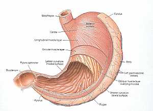
Stomach anatomy
09/12/2009 06:08:00 ص
this image shows the anatomy of the stomach showing its main features and parts.in this images we see the wall of the stomach being removed from the anterior portion to display the contents of the sto... More Details
Cranial nerves anatomy
05/11/2009 04:03:00 ص
this image shows the all cranial nerves and displaying their effector organs showing: 1. Olfactory nerve I 2. Optic nerve II 3. Occulomotor nerve III 4. Trochlear nerve IV 5. Trigeminal nerve V 6. Ab... More Details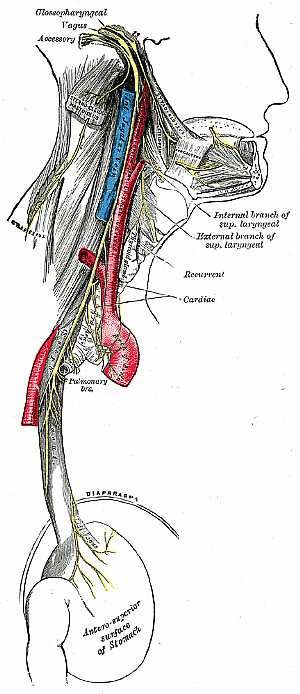
Vagus nerve anatomy
30/10/2009 03:55:00 م
this image shows the course of the vagus nerve in relation to the surrounding structures showing: 1. glossopharyngeal nerve 2. vagus nerve 3. accessory nerve 4. internal jugular nerve 5. common carot... More Details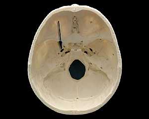
Skull Anatomy
19/10/2009 03:02:24 م
In This Section you will find detailed different Photos and images about the anatomy of the Skull bone including its surface , attachments related structures many more Items about the Skull anatomy... More DetailsCranial nerves anatomy
22/10/2009 01:51:53 م
In This Section you will find detailed different Photos and images about the anatomy of the Cranial Nerves including Their types , Fascial nerve anatomy , trigeminal nerve anatomy , vagus nerve anatom... More Details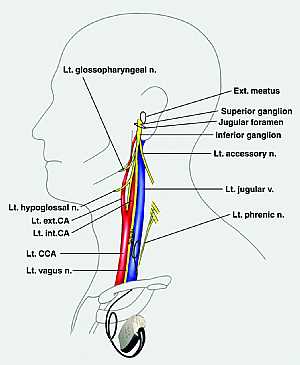
Glossopharybgeal nerve anatomy
29/10/2009 03:23:00 م
this is an image of the lateral side of the neck displaying the left glossopharyngeal nerve related to the surrounding structures showing: 1. left glossopharyngeal nerve 2. external auditory meatus 3... More Details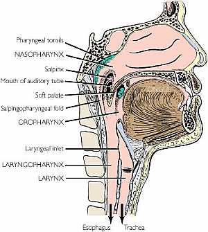
The pharynx anatomy
12/10/2009 04:24:00 ص
this is a longitudinal section in the head and the neck showing the structures of the pharynx ( nasophrynx,oropharynx and laryngopharynx) showing: 1. pharyngeal tonsils 2. nasopharynx 3. salpinx 4. op... More Details
Head Anatomy
19/10/2009 03:02:56 م
In This Section you will find detailed different Photos and images about the anatomy of the Head including The Face , Muscles of the face , bones of the face and vessles many more Items about the Skul... More Details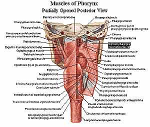
Pharynx anatomy
11/10/2009 04:13:00 م
this is posterior view of the muscles of the pharynx ( which is just the beginning of both the trachea and esophagus) showing: 1. pharyngobasilar fascia 2. accessory muscle bundle 3. digastric m. 4. s... More Details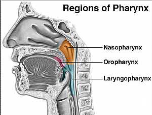
Pharynx anatomy
12/10/2009 04:34:00 ص
this image explains the position of the three parts of the pharynx showing: 1. nasopharynx 2. oropharynx 3. laryngopharynx... More Details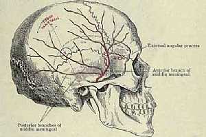
Blood supply of the exterior of the skull
09/10/2009 03:39:00 م
this image shows the middle meningeal artery (its course and branches) that artery supplies that exterior aspect of the skull it is also shows the common points for injury of that artery causing hemor... More Detailsorthopaedic joint assessment centr dr mcmahon
, , , , , , ,anatomi ligamen panggul wanita
, ,abdomen sans preparation normale
, , , , , ,world conferences on urine therapy
, , , , , , , , , ,© Copyright 2001-2022 eDoctorOnline.com