mss
humerus, nasal cavity, midbrain, neck muscles, stomach, humerus bone, FEMUR, eye anatomy, stomach anatomy, pelvic girdle, leprosy, visual pathway, eye diagram, lung anatomy, skeletal system, foramen rotundum, mouth anatomy, coronary arteries, muscles of the neck, nose anatomy,
The following are the result pages for the searched keyowrd: mss

Get Pregnant After 35 (and 40!)
09/06/2006
Assess the odds, up your chances, and glean advice from a midlife mom When I was 35, unmarried but still hoping for children someday, I asked my doctor if I should be concerned about my chances of get... More Details
Headaches can be relieved with calcium and vitamin D
19/10/2009
Headaches are one of the most common health complaints of adults. There are three types of headaches; tension, migraine, and cluster. The tension headache is the most common and often results from mus... More DetailsFat Burning Foods For A Flat Stomach
02/08/2009
Ok so your diet is working well and your workouts are going great - but you still have a little spot of fat on your stomach that just won't budge. Have you ever thought of looking into foods that ... More DetailsUse of Statistics in Hospital and Healthcare Organizations
22/01/2010
Now-a-days healthcare organizations are very much competitive. And they want to lead in all the areas of improvement. But sometimes they have to face big challenges for solutions to analytical problem... More DetailsUNICEF RECURITMENT
10/06/2009 06:13:00 ص
UNICEF IS CURRENTLY RECURITING NURSES,DOCTORS AND EVERY ONE UNDER MEDICAL FIELD.WE ARE JOB APPLICATION AGENT FOR THEM IF INTERESTED KINDLY SEND US A MAIL. JOB VERY LUCREATIVE WITH HIGY PAID SALARY... More Details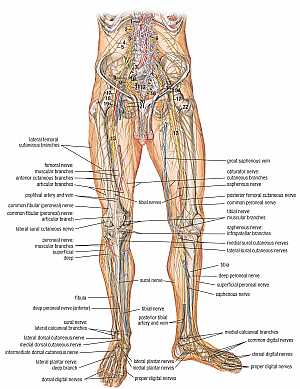
Leg nerves and vessels
15/07/2010 03:22:46 م
In This Section you will find detailed different Photos and images about the anatomy of the Leg nerves and vessels including parts , related structures and many more Items about the Leg nerves and ves... More DetailsMiller Fisher Syndrome: A Case Report
21/08/2010
Miller Fisher Syndrome is a rare variant of Guillain-Barre syndrome resulting in a post-infectious neuropathy that affects 0.1 per 100,000 worldwide annually. Miller Fisher syndrome presents with a tr... More Details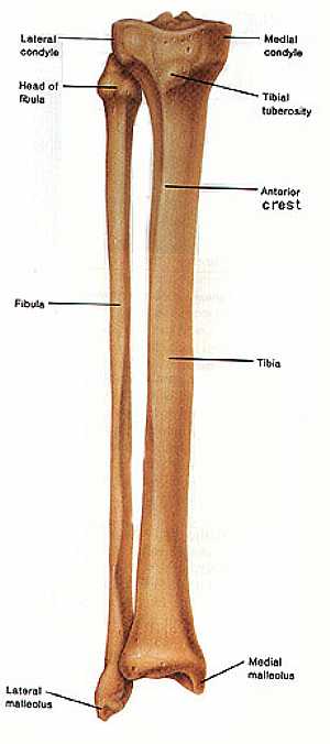
Fibula and Tibia bones anatomy
04/12/2009 07:43:00 ص
this image shows the anatomy of the fibula and tibia bones (the bones of the leg) in relation to each other ,displaying the different features and parts of them ,fibula (on the left) and the tibia (on... More Details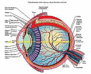
Eye anatomy
11/10/2009 04:00:00 م
this diagram details the different parts and structures of the human eye showing: 1. conjunctiva 2. ora serrata 3. cilliary body 4. aqueous 5. iris 6. ant. chamber 7. cornea 8. pupil 9. lens 10. post.... More Details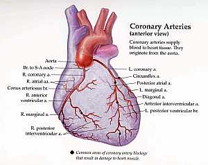
Coronary arteries anatomy
20/12/2009 02:25:00 م
this image shows the coronary arteries of the heart ( the arteries that supply the heart muscle with oxygen and nutrients) From anterior view .these arteries when occluded paretially or completely it ... More DetailsHow long will i live with mesothelioma?
07/10/2010
Mesothelioma (From greek, Meso=Middle and Thelio=Tunic, Clothing), is a malignant tumor of the serous membranes of the body. This membranes serve as lining between external and internal parts of the b... More DetailsIntroduction to Diabetes Mellitus - Diabetes 101
30/07/2009
Diabetes Mellitus (DM) is the term used to describe a group of similar disorders where affected individuals have too much glucose, or sugar, in their blood. The medical term for this is hyperglycemia.... More Details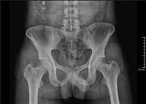
Male Pelvic X-Ray
09/01/2012 12:25:00 م
This is a X-Ray Image of Male Pelvis which is usually characterized by being Narrower and longer the female pelvis. Showing : 1. 2 Iliac Bones 2. 2 Sacroiliac joints 3. Sacrum 4. Lumbar Vertebrae 5. ... More Details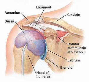
Shoulder joint anatomy
18/12/2009 07:48:00 ص
this image shows the anatomy of the shoulder joint from anterior view displaying the bones , ligaments and bursa of that joint. showing: 1. Acromion process of the scapula 2. Bursa of the shoulder jo... More Details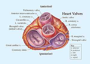
heart valve anatomy
07/01/2010 02:27:00 م
This image is a horizontal cut section of the heart at the level of the heart valves showing the four main valves of the heart with the related structures and vessels showing: 1. Pulmonary valve 2. A... More Details
WHO & FDA Release News
16/01/2010
Stay updated with the daily news and events in the medical field , the new releases of the WHO , FDA and the new breakthroughs in field of medicine and drugs ... More Details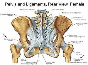
Bones and ligaments of the FEMALE Pelvis
22/11/2009 06:02:00 ص
this image shows the posterior "back" view of the female pelvic brim (the bones and ligaments that forms the pelvic region in the female) showing: 1. supraspinous ligament 2. posterior sacroi... More Details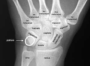
Wrist Joint X-Ray
09/01/2012 12:49:00 م
This is a X-Ray image of the Wrist joint showing the Wrist from antero-posterior view Showing : 1. Metacarpal bones 2. Trapezoid 3. Trapezium 4. Capitate 5. Hamate 6. Triquetrum 7. Lunate 8. Scaphoid... More Details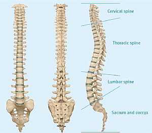
Spinal cord anatomy
22/10/2009 01:52:10 م
In This Section you will find detailed different Photos and images about the anatomy of the Spinal cord including its surface , parts , related structures , Functions of the spinal; cord , Spinal nerv... More Details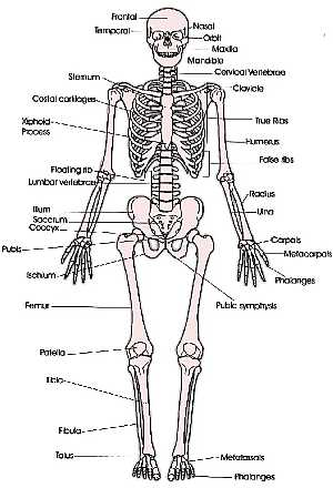
Anterior Skeleton (skeletal system)
05/03/2009 03:11:00 م
this image shows the skeleton system of our body (the bones that forms and supports our body) showing: 1. frontal bone of the skull 2. temporal bone of the skull 3. sternum 4. costal cartilages 5. xi... More DetailsHow does the body maintain normal blood pressure?
18/04/2006
The body has mechanisms to alter or maintain blood pressure and the flow of blood. There are sensors in the walls of the arteries and heart that sense blood pressure.... More Details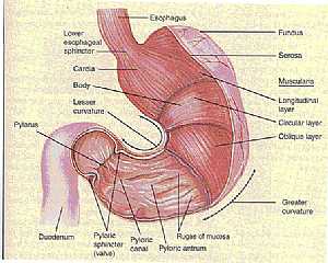
Stomach anatomy
09/12/2009 06:09:00 ص
this image shows the anatomy of the stomach showing its main features and parts.in this images we see the wall of the stomach being removed from the anterior portion to display the contents of the sto... More DetailsArticles for Physicians - Articles by Doctors
27/04/2006
This section contains articles and case studies. It is forwarded and designated to doctors, medical professionals and medical students who are seeking information about deferent medical fields and top... More Details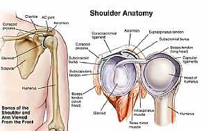
Shoulder joint anatomy
18/12/2009 07:45:00 ص
This image shows the anatomy of the shoulder joint from anterior view (on the left) and an open view (on the right) showing: 1. Supraspinatus tendon 2. Subacromial bursa 3. Biceps tendon (long head) ... More DetailsA Case of Abruptio Placentae with a History of H1N1 Influenza Infection
14/11/2009
Y.S. was a 19 y/o Hispanic female who presented to the labor and delivery ward complaining of abdominal pain and vaginal bleeding. The patient was G1 P0, and at 33+1 weeks. Estimated date of confineme... More Details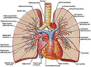
Heart and lung anatomy
07/12/2009 06:43:00 ص
this image shows the anatomy of the heart and the lungs in relation to each other displaying their different parts and features and the vessels of the heart and their relation to the lungs showing: 1... More Details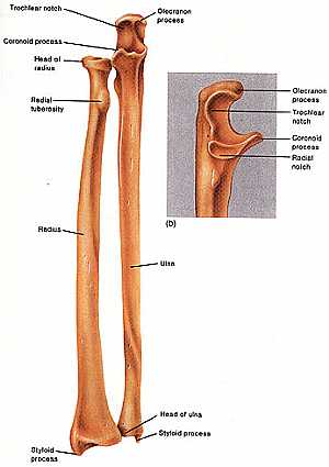
Radius an Ulna bones
04/12/2009 07:11:00 ص
this image shows the anatomy of the ulna and radius bones displaying their different features and parts of them showing: "ulna" 1. olecranon process 2. trochlear notch 3. head of the ulna 4. ... More Details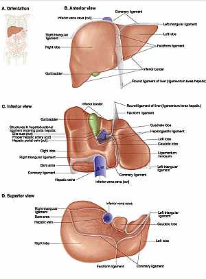
LIVER ANATOMY
05/03/2009 04:47:00 م
Pictures from Lippincott Williams & Wilkins Atlas of Anatomy, 1st Edition. 2008 this image shows the liver anatomy from ant. , superior and inferior views showing: "anterior" 1. coronary lig... More Details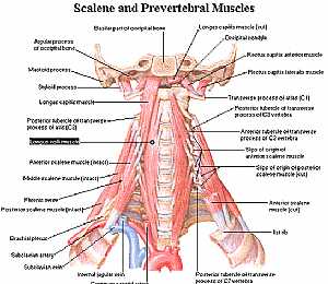
Neck Anatomy
11/10/2009 04:06:00 م
this is a back view of the muscles of the neck showing: 1. longus capitus m. 2. longus coli 3. ant. scalene m. 4. middle scalene m. 5. phrenic nerve 6. post. scalene m. 7. brachial plexus 8. subclavia... More Details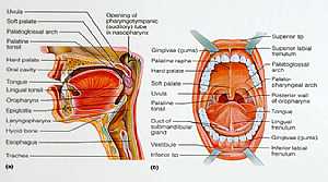
Oral Cavity
11/10/2009 04:09:00 م
the image on the right: is a detailed image of the structures of the mouth the image on the left: cut section in the head showing the structures of the mouth and oropharynx ( just after the mouth area... More Details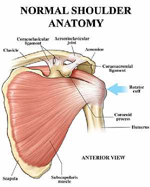
Shoulder joint anatomy
18/12/2009 07:18:00 ص
this image shows the anatomy of the shoulder joint from anterior view displaying the bones, ligaments and muscles in relation to each other. showing: 1. Clavicle bone 2. Coracoclavicular ligament 3. ... More Details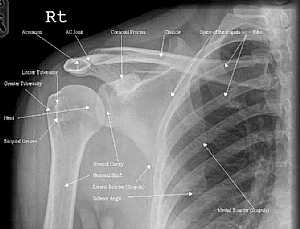
Shoulder Joint X-Ray
09/01/2012 12:55:00 م
This is a X-Ray image of the Shoulder joint showing the shoulder from antero-posterior view Showing : 1. Medial Border of Scapula 2. Ribs 3. Clavicle 4. Coronoid Process 5. Acromio-Clavicular Joint 6... More Details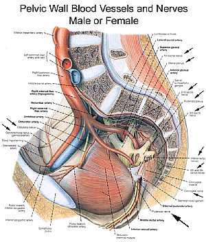
Pelvic nerves and vessels
13/11/2009 07:16:00 ص
this image is a side view of the vertically sectioned pelvis showing the arteries and nerves of the pelvic region showing: 1. inferior mesenteric artery 2. left common iliac artery 3. left common ili... More Details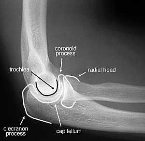
Elbow Joint X-Ray Lateral View
09/01/2012 12:51:00 م
This is a X-Ray image of the Elbow joint showing the Elbow from Lateral view. Showing : 1. Coronoid Process 2. Radial Head 3. Capitullum 4. Olecranon Process 5. Trochlea... More Details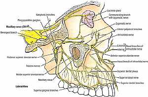
Trigeminal nerve anatomy
28/10/2009 01:55:00 ص
this images illustrates the different branches of the trigeminal nerve in the face in relation to each other [focusing on the maxillary division] showing: 1. maxillary nerve 2. meningeal branch 3. po... More Details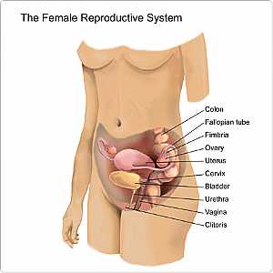
Female Reproductive Organs - Front View (Reproductive system of female)
27/04/2006
The female reproductive organs include the vagina (a muscular passage that connects the cervix with the external genital organs - one of which is a sensitive mound of tissue called the clitoris), the ... More Details
Skull Anatomy
19/10/2009 03:02:24 م
In This Section you will find detailed different Photos and images about the anatomy of the Skull bone including its surface , attachments related structures many more Items about the Skull anatomy... More DetailsWhen is the best time to get pregnant?
21/04/2006
The best or most fertile time to get pregnant is the period of ovulation in your menstrual cycle. This is the time when following an LH surge, a mature ovum is released into the uterus from the follic... More DetailsOpening in Saudi - Head Nurse
01/05/2009 12:18:00 م
Opening in one of the largest Hospital in Saudi Arabia Head Nurse Department : Medical Operation Supervisory responsibility : • Responsible to : Chief nurse • Responsible for : nurses & medical ... More DetailsMedical Jobs
02/03/2009 05:26:41 م
A free job service, where medical related job seekers and employers can post their information and search within job listings. Doctors, Physicians, Nurses, Pharmacist and other Healthcare job seekers... More Details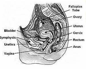
Vagina anatomy
13/11/2009 06:27:00 ص
this image shows the vagina of the female pelvis in relation to the surrounding organs this is a side view of a vertically sectioned pelvis showing: 1. fallopian tube 2. ovary 3. uterus 4. cervix 5. r... More Details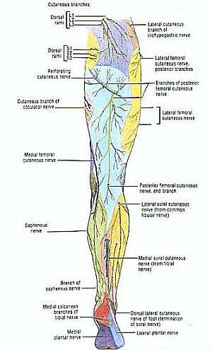
Nerves of the lower limb anatomy
22/11/2009 06:56:00 ص
this image shows the cutaneous nerve supply of the lower limb ( the nerves that supply the skin of the lower limb) "from posterior view" showing: 1. dorsal rami of (L1-L2-L3) 2. dorsal rami o... More DetailsNerves of the lower limb anatomy
24/11/2009 06:08:00 ص
this image shows the nerves of the lower limb showing their course , relation , branches and distribution (from anterior view) showing: "numbers" abdomen: 1. hepatic plexus 2. L1 nerve 3. L2... More Details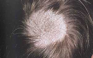
Tinea Capitis (gray Patch)
31/07/2011 07:28:00 م
Tinea Capitis (gray Patch) The lesion is a small, round, elevated patches, hyperkeratotic , scaling of the scalp, and dry and brittle hair ( the hair shafts are broken just above the surface)... More Details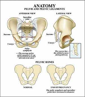
Pelvic bone anatomy
13/11/2009 06:32:00 ص
this image shows the pelvic bone that supports the pelvic region from anterior and lateral view showing: 1. sacroiliac joint 2. sacrum 3. coccyx 4. symphysis pubis 5. hib bone... More Details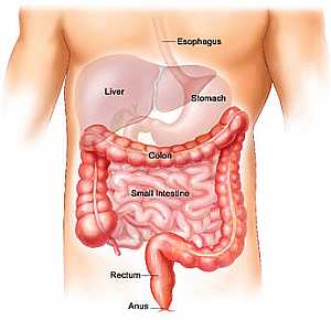
Small and Large intestine anatomy
09/12/2009 05:48:00 ص
this image shows the anatomy of the GIT from the small intestine to the anus.displaying their position and relation to each other and to the rest of the body. showing: 1. esophagus 2. Liver 3. Stomac... More Details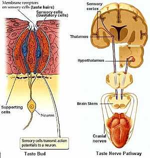
Taste sensation pathway
03/11/2009 02:31:00 م
this image shows the pathway of the taste sensation from the tongue to the central nervous system the left image shows the histology of the taste bud ( the taste receptor of the tongue) showing: 1. t... More Details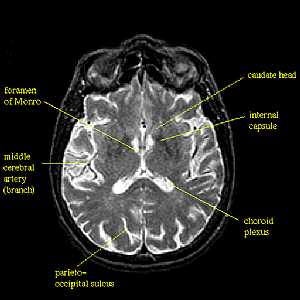
Brain MRI
06/03/2012 11:47:00 ص
This image shows a transverse section of the brain using MRI technique (notice that the bone here is black in contrast to CT in which the bone is white) This technique shows the details of thee soft t... More Details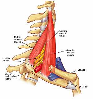
Neck Muscles
06/07/2012 09:05:00 م
1. Middle scalene muscle 2. Scalene Muscle triangle 3. Anterior scalene Muscle 4. Brachial Plexus 5. Axillary Artery... More Details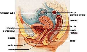
Female pelvic anatomy
13/11/2009 07:06:00 ص
this image shows lateral view of the vertically sectioned pelvis of female showing: 1. fallopian tube 2. urinary bladder 3. pubic bone 4. clitoris 5. urethra 6. vagina 7. anus 8. rectum 9. cervix 10.... More DetailsThe Gestational Surrogacy Process,Affordable surrogacy option.
21/03/2011
Going for surrogacy is a very big decision. It involves lot of ethical, moral, and also financial issues. This is the reason you need to be very careful while taking a step ahead in choosing surrogate... More Details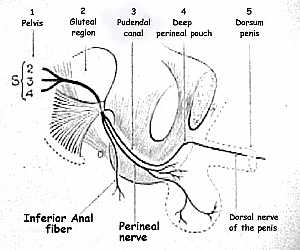
Pudendal nerve anatomy
13/11/2009 06:21:00 ص
this image shows the pathway of the pudendal nerve in the abdominal region showing: 1. pudendal nerve 2. sacral spinal nerves (S2--S4) 3. gluteal region 4. pudendal canal 5. deep perineal pouch 6. do... More Details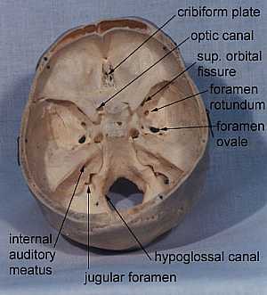
the main openings in the skull
10/10/2009 03:33:00 م
FORAMEN : CN cribiform plate I optic canal II sup. orbital fissure III, IV, Va, VI foramen rotundum Vb foramen ovale Vc int. auditory meatus VII, VIII jugular foramen IX, X, XI hypoglossal ca... More Details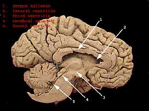
Brain ventricles
13/10/2009 01:20:00 ص
this section that shows the medial wall of the cerebral hemisphere shows the main four ventricles of the brain showing: 1. corpus callosum 2. lateral ventricle 3. cerebral aquiduct 4. fourth ventricle... More Details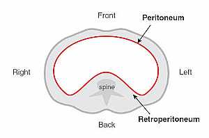
Peritoneum anatomy
12/11/2009 02:00:00 م
this image shows what is called the peritoneum of the abdomen (a membranous sac that is filled with fluid and surrounds the abdominal organs) this image is from superior view "this cut level there... More Details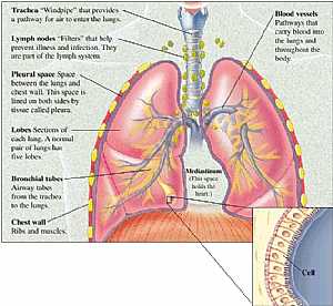
Respiratory system (chest anatomy)
07/12/2009 06:15:00 ص
this image shows the respiratory system anatomy from the trachea to the lungs.Displaying the different parts and relations of the different organs in that system showing: 1. Trachea 2. Lymph nodes 3.... More Details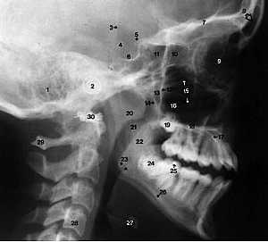
Skull X-Ray Lateral View
09/01/2012 11:35:00 ص
This is an X-Ray image of the Skull taken from a Lateral View showing the Skull From the Side. Showing: 1. Mastoid air cells 2. Ear rods 3. Anterior Border of Head Positioner 4. Posterior Clinoid Pro... More Details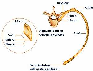
Typical rib anatomy
06/12/2009 05:41:00 ص
this image shows the anatomy of the typical rib in position with the vertebra displaying its different features and the relation of the groove ( the VAN "vein , artery,nerve") showing: 1. ver... More DetailsLaser Surgery for Ear Infections (OtoLAM)
07/06/2006
I am looking for help with my four-year-old son. From the age of six months, he has had tubes in his ears for chronic ear infections and hearing impairment.... More Details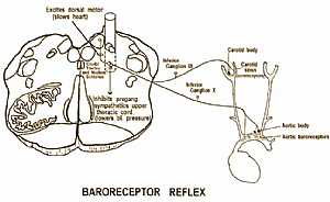
Baroreceptor reflex pathway
02/11/2009 02:20:00 م
this image shows the pathway of the baroreceptor reflex "the reflex that shares in the control of normal blood pressure" showing: 1. aortic baroreceptor 2. aortic body 3. carotid sinus 4. car... More Details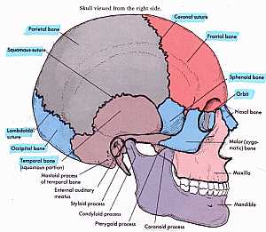
Skull Anatomy
10/10/2009 04:28:00 م
this shows the names of the bones that forms the side of the skull showing: 1. coronal suture 2. frontal bone 3. sphenoid bone 4. orbital bone 5. nasal bone 6. zygomatic bone 7. maxillary bone 8. mand... More DetailsCarrying Twins!
21/04/2006
The chances of a pregnant woman having twins or a multiple birth have never been higher. According to the National Center for Health Statistics, about one in every 35 babies born is a multiple (twin, ... More DetailsA Myth-Proof Guide to Getting Pregnant
21/04/2006
When it comes to getting pregnant, simply having made a baby does not make you an expert. Yet once you and your partner announce that you’re trying to conceive, everyone from your jeweler to your sist... More Details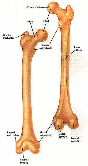
Femur bone anatomy
04/12/2009 07:30:00 ص
this image shows the anatomy of the femur bone from anterior and posterior view displaying its different features and parts showing: 1. fovea capitis 2. head of the femur 3. neck of the femur 4. less... More DetailsIndia Surrogacy: Real successes with affordable costs
04/05/2011
Are you looking out for some good and affordable surrogates who can lend you their womb? Well if this is the case then you can think of hunting for some good options on the web. There are some great S... More DetailsHow to get Pregnant Faster – Top Ten Tips
21/04/2006
simple tens tips on how to get pregnant Faster...... More Details7 Ways To Save Your Eyesight
18/04/2006
How much time do you spend in front of the computer? While eDiets members know first-hand many of the benefits that being online can provide, extensive computer work can take its toll on your eyes.... More DetailsHow I Treat Colds and Flu ?
23/05/2006
Colds and flu are probably the most commonly seen conditions by physicians in general or family practice.... More DetailsA Quick Guide On How To Get Pregnant
09/06/2006
Congratulations! So you’ve decided to take the big step into becoming a parent. While it is one of the most important things a person can do in their life, it is also the most challenging and the firs... More Details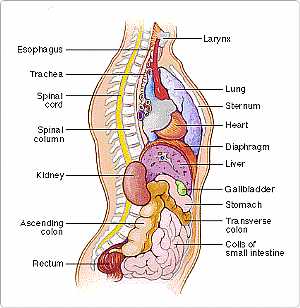
Body/Torso -- Side View (Body anatomy)
27/04/2006
Your torso consists of two parts — the chest and the abdomen. The chest contains your heart and lungs; your abdomen contains the digestive and urinary systems. Your chest and abdomen are separated by ... More Details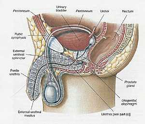
Pelvis Anatomy
06/11/2009 01:06:01 م
In This Section you will find detailed different Sections about the different organs and structures in the region of the Pelvis including Male pelvis anatomy , Female pelvis anatomy , pelvic girdle an... More Details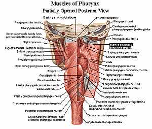
Pharynx anatomy
11/10/2009 04:13:00 م
this is posterior view of the muscles of the pharynx ( which is just the beginning of both the trachea and esophagus) showing: 1. pharyngobasilar fascia 2. accessory muscle bundle 3. digastric m. 4. s... More Details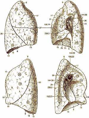
Lung anatomy (lung lobe anatomy )
09/11/2009 04:10:00 م
this image shows the anatomy of the surface of the lungs (right and left ) (medial and lateral surfaces) showing: 8. Base of lung 9. Apex of lung 10. Costal surface Facing ribs 11. Medial surface... More DetailsMale pelvis anatomy
10/12/2009 09:19:00 ص
this image shows the anatomy of the male pelvis in longitudinal section view the median line of the male pelvis.showing the male pelvic organs and structures in relation to each other. showing: 1. Pe... More Details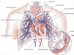
Lung anatomy
09/11/2009 04:19:00 م
this image shows the area of exchange of gases between the blood and the air in the lungs and the blood circulation in the thorax region showing: 1. superior vena cava 2. inferior vena cava 3. heart 4... More Details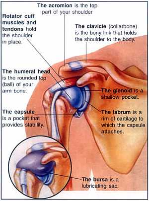
Shoulder joint anatomy
18/12/2009 07:34:00 ص
this image shows the shoulder joint from anterior view showing the bones and ligaments of the joint.also we can see what is called the synovial membrane of the joint ( the membrane that surrounds the ... More DetailsUnderstanding Your Heart Rate
05/05/2006
Your heart is a muscle that is located on the left side of your chest and is about the size of your fist. It sends blood thoughout your body, providing it with the proper nutrients and oxygen that it ... More Details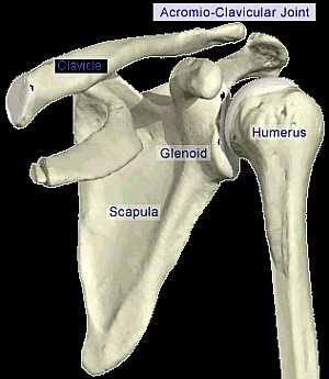
Acromio-Clavicular joint
18/12/2009 06:58:00 ص
this image shows the anatomy of the bones forming the shoulder joint from anterior view.displaying the acromio-clavicular joint(the joint between the acromion process of the scapula and the head of th... More Details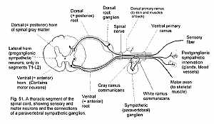
Spinal nerve connections
01/11/2009 02:42:00 م
this image shows all the connections between the spinal cord (spinal nerve) and the effector organs showing: 1. dorsal horn of spinal cord 2. lateral horn of spinal cord 3. ventral horn of spinal cor... More Details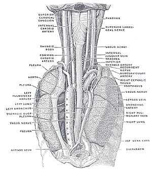
Vagus nerve anatomy
30/10/2009 04:02:00 م
this image shows the vagus nerve at the thorax showing: 1. aorta 2. left pulmonary artery 3. left lung 4. left bronchus 5. vagus nerve 6. pleura 7. azygot vein 8. inferior vena cava 9. aortic arch 10... More DetailsDiabetes Symptoms, When to See the Doctor?
18/04/2006
The cases of diabetes are growing in the United States today. This is due in part to our aging population as well as to our growing waistlines.... More DetailsCosmetic Surgery: Tummy Tuck, Arm and Thighs Lift and of course the Full Body Lift.
16/04/2006
With the rampant and almost epidemic manifestation of obesity nowadays it is not uncommon for the overweight person to have to lose a massive amount of weight.... More Details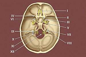
Base of the skull
09/10/2009 03:34:00 م
this image shows the exit of the twelve cranial nerves from the base of the skull * I. Olfactory nerve * II. Optic nerve * III. Oculomotor nerve * IV. Trochlear nerve * V. Trigeminal nerve * VI.... More DetailsNitroglycerin translingual spray - Drug Information
contains Nitroglycerin translingual spray drug information and providing Nitroglycerin translingual spray drug indication, contraindication, special concerns, side effects, overdose management and dosage... More Details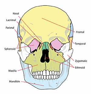
Skull Anatomy
10/10/2009 04:26:00 م
this image shows the names of the bones that forms the front of the skull showing: 1. nasal bone 2. lacrimal bone 3. temporal bone 4. zygomatic bone 5. ethmoid bone 6. mandible bone 7. sphenoid bone 8... More Details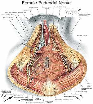
Female pelvic nerves and vessels
13/11/2009 06:54:00 ص
this image shows the pelvic arteries , veins and nerves in relation to each other and to the surrounding structures in the female pelvis region showing: 1. ischial tuberosity 2. superfaicial transver... More Details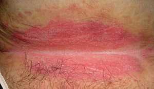
Intertrigo
31/07/2011 07:12:00 م
Intertrigo An intertrigo usually develops from the chafing of warm, moist skin in the areas of the inner thighs and genitalia, the armpits, under the breasts, the underside of the belly, behind the e... More Details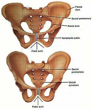
Pelvic girdle anatomy
04/12/2009 07:39:00 ص
this image shows the bones that forms the pelvic girdle (the hib bone,the sacrum and the symphysis pubis) in relation to each other showing: 1. ilium bone 2. pelvic brim of the pelvis 3. symphysis pu... More Details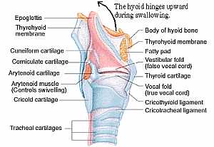
Anatomy of the larynx
11/10/2009 03:44:00 م
this is the structure of the larynx ( the throat area ) showing: 1. epiglottis 2. thyroid membrane 3. cuneiform cartilage 4. corniculate cartilage 5. arytenoid cartilage 6. arytenoid m. 7. cricoid car... More DetailsMedical Insurance - Health Insurance Coverage & Plans Explained
11/06/2006
If you're trying to decide between health insurance plans, you'll find that there are several different kinds from which to choose. It's important that you weigh all your options carefully... More Detailsorthopaedic joint assessment centr dr mcmahon
, , , , , , ,anatomi ligamen panggul wanita
, ,abdomen sans preparation normale
, , , , , ,world conferences on urine therapy
, , , , , , , , , ,© Copyright 2001-2022 eDoctorOnline.com