pelvis bone anatomy
pelvic girdle, pelvic girdle, pelvic ligaments, Pelvis, pelvis, pelvic ligaments, pelvis, pelvic ligaments, female pelvic anatomy, bones of the pelvis, pelvic anatomy, ligaments of the pelvis, bones of the pelvis, ligaments of the pelvis, interosseous sacroiliac ligament, pubic arch, sacrotuberous ligament, pelvic, pelvic, sacrospinous ligament,
The following are the result pages for the searched keyowrd: pelvis bone anatomy
Pelvic bone anatomy
13/11/2009 06:32:00 ص
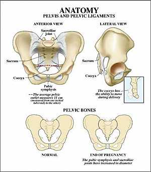
this image shows the pelvic bone that supports the pelvic region from anterior and lateral view showing: 1. sacroiliac joint 2. sacrum 3. coccyx 4. symphysis pubis 5. hib bone... More Details
Bones and ligaments of the FEMALE Pelvis
22/11/2009 06:02:00 ص
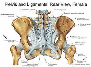
this image shows the posterior "back" view of the female pelvic brim (the bones and ligaments that forms the pelvic region in the female) showing: 1. supraspinous ligament 2. posterior sacroi... More Details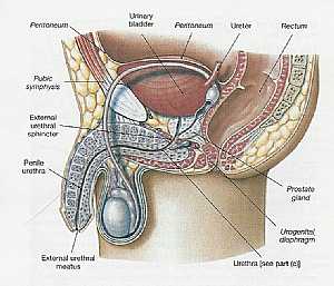
Pelvis Anatomy
06/11/2009 01:06:01 م
In This Section you will find detailed different Sections about the different organs and structures in the region of the Pelvis including Male pelvis anatomy , Female pelvis anatomy , pelvic girdle an... More Details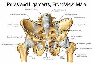
Bones and ligaments of the MALE Pelvis
22/11/2009 05:57:00 ص
this image shows the anterior view of the male pelvic brim ( the bones and ligaments that forms and supports the pelvic region) showing: 1. 4th lumbar vertebra 2. anterior sacroiliac ligament 3. ante... More Details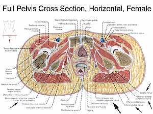
Female pelvic anatomy
13/11/2009 07:31:00 ص
this image is a horizontal section in a female pelvis shows the different pelvic organs and structures in relation to each other showing: 1. iliacus muscle 2. sartorius muscle 3. rectus femoris muscl... More DetailsBones and ligaments of the MALE Pelvis
22/11/2009 05:57:00 ص
this image shows the anterior view of the male pelvic brim ( the bones and ligaments that forms and supports the pelvic region) showing: 1. 4th lumbar vertebra 2. anterior sacroiliac ligament 3. ante... More Details
Pelvic bone and ligaments anatomy
13/11/2009 07:39:00 ص
this image shows the boundaries of the pelvic area formed of the pelvic bones and ligaments showing: 1. iliolumbar ligament 2. iliac crest 3. fifth lumbar vertebra 4. sacral promontory 5. anterior su... More Details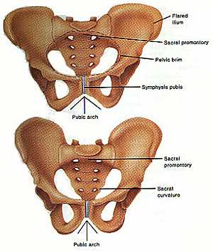
Pelvic girdle anatomy
04/12/2009 07:39:00 ص
this image shows the bones that forms the pelvic girdle (the hib bone,the sacrum and the symphysis pubis) in relation to each other showing: 1. ilium bone 2. pelvic brim of the pelvis 3. symphysis pu... More Details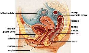
Female pelvic anatomy
13/11/2009 07:06:00 ص
this image shows lateral view of the vertically sectioned pelvis of female showing: 1. fallopian tube 2. urinary bladder 3. pubic bone 4. clitoris 5. urethra 6. vagina 7. anus 8. rectum 9. cervix 10.... More Details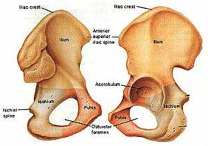
Hib bone anatomy
04/12/2009 07:22:00 ص
this image shows the anatomy of the hib bone from anterio-lateral (on the left) and from posterio-lateral view(on the right) showing: 1. iliac crest 2. ilium bone 3. ischial bone 4. pubis 5. obturato... More Details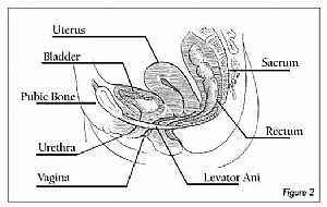
Female pelvis anatomy
13/11/2009 06:19:00 ص
this image shows a side view of the female pelvis anatomy sectioned vertically showing: 1. uterus 2. urinary bladder 3. pelvic bone 4. urethra 5. vagina 6. levator ani muscle 7. rectum 8. sacrum bone... More DetailsPelvic girdle anatomy
04/12/2009 07:39:00 ص
this image shows the bones that forms the pelvic girdle (the hib bone,the sacrum and the symphysis pubis) in relation to each other showing: 1. ilium bone 2. pelvic brim of the pelvis 3. symphysis pu... More Details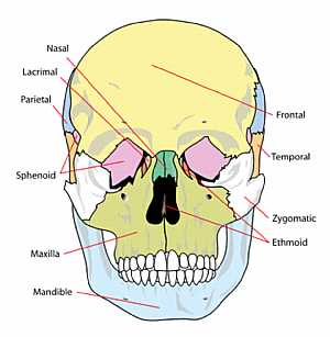
Skull Anatomy
10/10/2009 04:26:00 م
this image shows the names of the bones that forms the front of the skull showing: 1. nasal bone 2. lacrimal bone 3. temporal bone 4. zygomatic bone 5. ethmoid bone 6. mandible bone 7. sphenoid bone 8... More DetailsPelvic bone and ligaments anatomy
13/11/2009 07:39:00 ص
this image shows the boundaries of the pelvic area formed of the pelvic bones and ligaments showing: 1. iliolumbar ligament 2. iliac crest 3. fifth lumbar vertebra 4. sacral promontory 5. anterior su... More Detailsorthopaedic joint assessment centr dr mcmahon
, , , , , , ,anatomi ligamen panggul wanita
, ,abdomen sans preparation normale
, , , , , ,world conferences on urine therapy
, , , , , , , , , ,© Copyright 2001-2022 eDoctorOnline.com