pudendal nerve anatomy
pelvic anatomy, anatomy, pelvic, uterus anatomy, round ligament, pelvic bone anatomy, female anatomy, female pelvic anatomy, male, PUDENDAL NERVE, uterosacral ligament, pelvic bone, pelvic nerves, round ligament of uterus, pudendal nerve, anatomy pelvis, pelvic anatomy, pelvis anatomy, pelvic region, pudendal canal,
The following are the result pages for the searched keyowrd: pudendal nerve anatomy
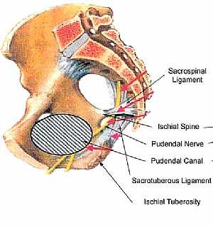
Pudendal nerve anatomy
13/11/2009 06:43:00 ص
this image shows the pudendal nerve in the pelvic showing its course ond relation to the surrounding structures showing: 1. sacrospinal ligament 2. iscial spine 3. pudendal nerve 4. pudendal canal 5.... More Details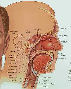
anatomy of the head and neck
09/10/2009 03:44:00 م
this image shows longitudinal section in the head and neck with their details (the frontal,nasal,oral and neck regions) showing: 1. nasolacrimal duct 2. sella turcica 3. sphenoid sinus 4. frontal sinu... More Details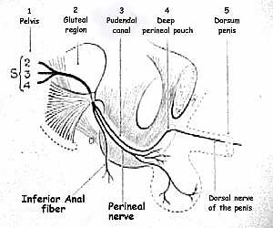
Pudendal nerve anatomy
13/11/2009 06:21:00 ص
this image shows the pathway of the pudendal nerve in the abdominal region showing: 1. pudendal nerve 2. sacral spinal nerves (S2--S4) 3. gluteal region 4. pudendal canal 5. deep perineal pouch 6. do... More Details
Pelvis Anatomy
06/11/2009 01:06:01 م
In This Section you will find detailed different Sections about the different organs and structures in the region of the Pelvis including Male pelvis anatomy , Female pelvis anatomy , pelvic girdle an... More Details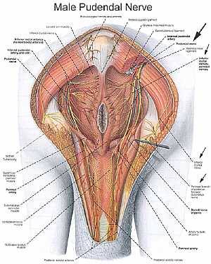
Male pelvic nerves and vessels
13/11/2009 07:01:00 ص
this image shows the pelvic arteries , veins and nerves in relation to each other and to the surrounding structures in the female pelvis region showing: 1. anococcygeal nerves 2. anococcygeal arterie... More Details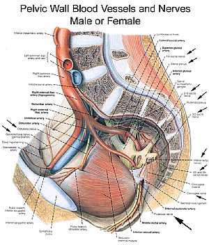
Pelvic nerves and vessels
13/11/2009 07:16:00 ص
this image is a side view of the vertically sectioned pelvis showing the arteries and nerves of the pelvic region showing: 1. inferior mesenteric artery 2. left common iliac artery 3. left common ili... More DetailsPudendal nerve anatomy
13/11/2009 06:21:00 ص
this image shows the pathway of the pudendal nerve in the abdominal region showing: 1. pudendal nerve 2. sacral spinal nerves (S2--S4) 3. gluteal region 4. pudendal canal 5. deep perineal pouch 6. do... More Details
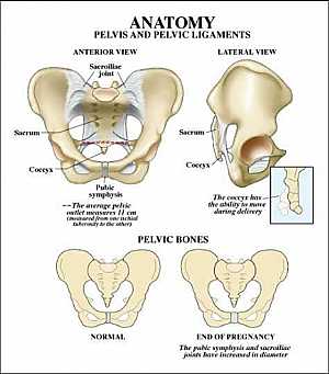
Pelvic bone anatomy
13/11/2009 06:32:00 ص
this image shows the pelvic bone that supports the pelvic region from anterior and lateral view showing: 1. sacroiliac joint 2. sacrum 3. coccyx 4. symphysis pubis 5. hib bone... More Details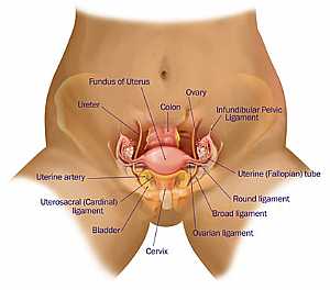
Female pelvic anatomy
13/11/2009 07:04:00 ص
this is an anterior view of the female reproductive system in the pelvic region showing: 1. fundus of the uterus 2. ureter 3. colon 4. ovary 5. infundibular pelvic ligament 6. uterine (fallopian ) tu... More Details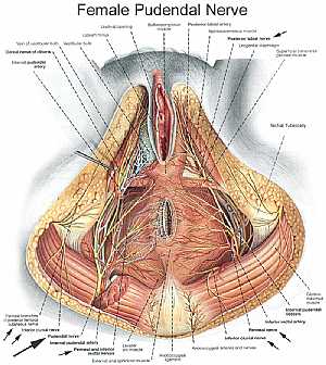
Female pelvic nerves and vessels
13/11/2009 06:54:00 ص
this image shows the pelvic arteries , veins and nerves in relation to each other and to the surrounding structures in the female pelvis region showing: 1. ischial tuberosity 2. superfaicial transver... More DetailsRelated Searches
loading...
loading...
Top Health & Medical Articles
New Articles
Most Read
آخر كلمات البحث
orthopaedic joint assessment centr dr mcmahon
, , , , , , ,anatomi ligamen panggul wanita
, ,abdomen sans preparation normale
, , , , , ,world conferences on urine therapy
, , , , , , , , , ,eDoctorOnline.com does not provide medical advice, diagnosis or treatment.
© Copyright 2001-2022 eDoctorOnline.com
© Copyright 2001-2022 eDoctorOnline.com