shoulder anatomy
neck muscles, neck anatomy, shoulder joint, muscles of the neck, trapezius muscle, sternocleidomastoid, shoulder joint, neck, thalamus, shoulder joint anatomy, shoulder joint anatomy, neck anatomy, trapezius, neck muscles anatomy, shoulder muscle anatomy, muscles in the neck, ear anatomy, anatomy of the neck, anatomy of neck, shoulder anatomy,
The following are the result pages for the searched keyowrd: shoulder anatomy
Shoulder joint anatomy
18/12/2009 07:34:00 ص
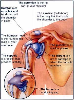
this image shows the shoulder joint from anterior view showing the bones and ligaments of the joint.also we can see what is called the synovial membrane of the joint ( the membrane that surrounds the ... More Details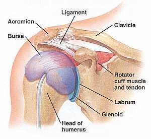
Shoulder joint anatomy
18/12/2009 07:48:00 ص
this image shows the anatomy of the shoulder joint from anterior view displaying the bones , ligaments and bursa of that joint. showing: 1. Acromion process of the scapula 2. Bursa of the shoulder jo... More Details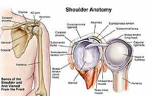
Shoulder joint anatomy
18/12/2009 07:45:00 ص
This image shows the anatomy of the shoulder joint from anterior view (on the left) and an open view (on the right) showing: 1. Supraspinatus tendon 2. Subacromial bursa 3. Biceps tendon (long head) ... More DetailsShoulder joint anatomy
18/12/2009 07:48:00 ص
this image shows the anatomy of the shoulder joint from anterior view displaying the bones , ligaments and bursa of that joint. showing: 1. Acromion process of the scapula 2. Bursa of the shoulder jo... More DetailsShoulder Anatomy
14/07/2010 11:39:39 ص
In This Section you will find detailed different Photos and images about the anatomy of the Shoulder including its surface , parts , related structures , Muscles of the shoulder , Brachial plexus , te... More Details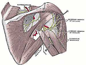
Shoulder joint anatomy
18/12/2009 07:38:00 ص
this image shows the anatomy of the shoulder joint from posterior view displaying the bones,ligaments,muscles,nerves and vessels of that region. (nerves are in yellow color,arteries are in red color) ... More Details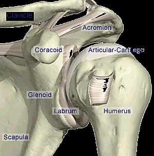
Shoulder joint anatomy
18/12/2009 07:07:00 ص
this image shows the anatomy of the shoulder joint from anterior view displaying the bones and the ligaments that forms that joint and their position in relation to each other. showing: 1. Clavicle b... More DetailsShoulder joint anatomy
18/12/2009 07:34:00 ص
this image shows the shoulder joint from anterior view showing the bones and ligaments of the joint.also we can see what is called the synovial membrane of the joint ( the membrane that surrounds the ... More Details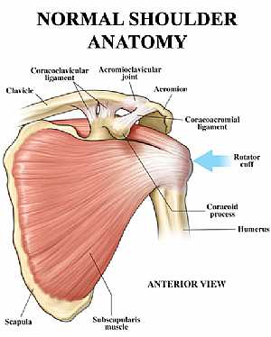
Shoulder joint anatomy
18/12/2009 07:18:00 ص
this image shows the anatomy of the shoulder joint from anterior view displaying the bones, ligaments and muscles in relation to each other. showing: 1. Clavicle bone 2. Coracoclavicular ligament 3. ... More Details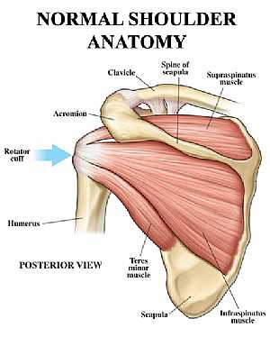
Shoulder joint anatomy
18/12/2009 07:20:00 ص
this image shows the anatomy of the shoulder joint from posterior view displaying the bones, tendons and muscles of the joint in relation to each other. showing: 1. Supraspinatus muscle 2. Spine of t... More Details
Shoulder joint anatomy
18/12/2009 07:45:00 ص
This image shows the anatomy of the shoulder joint from anterior view (on the left) and an open view (on the right) showing: 1. Supraspinatus tendon 2. Subacromial bursa 3. Biceps tendon (long head) ... More DetailsShoulder joint anatomy
18/12/2009 07:20:00 ص
this image shows the anatomy of the shoulder joint from posterior view displaying the bones, tendons and muscles of the joint in relation to each other. showing: 1. Supraspinatus muscle 2. Spine of t... More DetailsShoulder joint anatomy
18/12/2009 07:07:00 ص
this image shows the anatomy of the shoulder joint from anterior view displaying the bones and the ligaments that forms that joint and their position in relation to each other. showing: 1. Clavicle b... More Details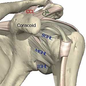
Shoulder joint anatomy
04/12/2009 07:36:00 ص
this image shows the anatomy of the shoulder joint diplaying the different ligaments that supports that joint and prevent it from dislocation showing: 1. CCL - coracoclavicular ligaments 2. CAL - cor... More Details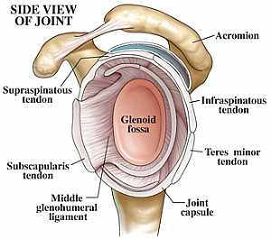
Shoulder joint anatomy
18/12/2009 07:30:00 ص
this image shows the anatomy of the shoulder joint from side lateral view.the humerus bone was removed showing the lateral side of the scapula bone.in this image we can see the glenoid process of the ... More Details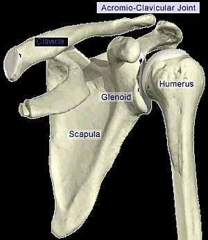
Acromio-Clavicular joint
18/12/2009 06:58:00 ص
this image shows the anatomy of the bones forming the shoulder joint from anterior view.displaying the acromio-clavicular joint(the joint between the acromion process of the scapula and the head of th... More Details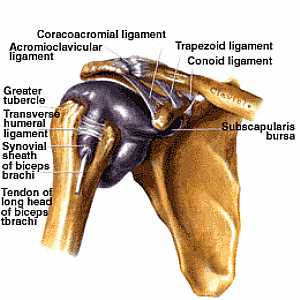
Shoulder joint anatomy
06/11/2009 03:00:00 م
this image shows the shoulder joint and the area of contact between the humerus and the scapula being covered with what is called the synovial sheath (to protect the joint) and displays the ligaments ... More DetailsShoulder joint anatomy
18/12/2009 07:38:00 ص
this image shows the anatomy of the shoulder joint from posterior view displaying the bones,ligaments,muscles,nerves and vessels of that region. (nerves are in yellow color,arteries are in red color) ... More Details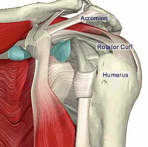
Shoulder joint anatomy
18/12/2009 07:15:00 ص
this image shows what is called the rotator cuff of the shoulder joint.the rotator cuff is a group of four muscles that surround the joint from four directions to support it and prevent it from disloc... More Details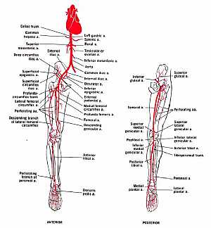
Arteries of the lower limb
22/11/2009 06:49:00 ص
this image shows the arteries that supply the lower limb showing their origin , course and branches (from anterior and posterior view) showing: "anterior view" 1. celiac artery 2. common hepa... More DetailsShoulder joint anatomy
04/12/2009 07:36:00 ص
this image shows the anatomy of the shoulder joint diplaying the different ligaments that supports that joint and prevent it from dislocation showing: 1. CCL - coracoclavicular ligaments 2. CAL - cor... More Details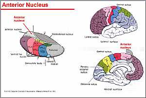
Thalamus anatomy
22/10/2009 02:21:00 م
this image shows the details of the thalamus and its nuclei showing: 1. anterior nuclei 2. ventral nuclei 3. geniculate body 4. lateral nuclei 5. medial nuclei 6. mediodorsal nucleus... More Details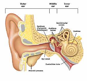
Ear anatomy
12/10/2009 04:42:00 ص
this shows the structure of the ear outer,middle and inner ear adjoined together showing: 1. mastoid process 2. ear canal 3. Eustachian tube 4. ear drum 5. auditory nerves 6. semicircular canals 7. co... More DetailsShoulder joint anatomy
18/12/2009 07:30:00 ص
this image shows the anatomy of the shoulder joint from side lateral view.the humerus bone was removed showing the lateral side of the scapula bone.in this image we can see the glenoid process of the ... More Details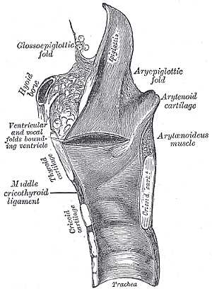
Larynx Anatomy
19/10/2009 03:06:37 م
In This Section you will find detailed different Photos and images about the anatomy of the Larynx including its surface , attachments , related structures , vocal cords and many more Items about the... More Details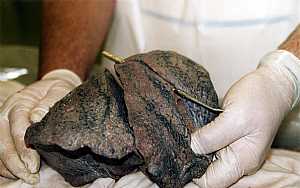
Lung anatomy
07/12/2009 06:31:00 ص
this image shows the anatomy of the right lung displaying its two main fissures the horizontal and the oblique one.they divide the right lung into three lobs "the upper lobe, the middle or the lat... More Details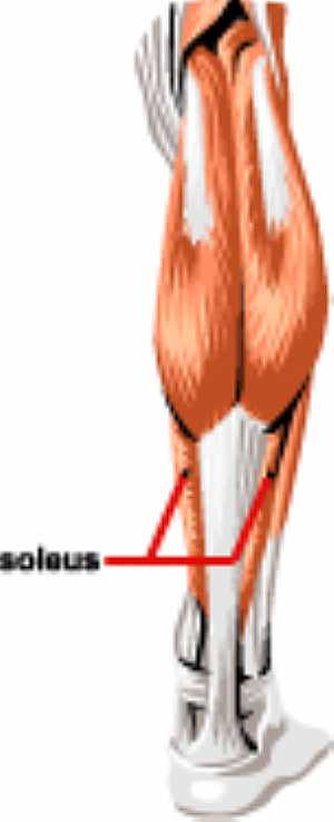
Leg muscles anatomy
15/07/2010 03:21:41 م
In This Section you will find detailed different Photos and images about the anatomy of the Leg muscles including its surface , parts , related structures , different muscles of the lower limbs and ma... More Details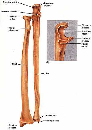
Forearm Anatomy
14/07/2010 11:41:20 ص
In This Section you will find detailed different Photos and images about the anatomy of the Forearm including its surface , parts , related structures and many more Items about the Forearm anatomy...... More Details
Atlas of Human Anatomy
27/04/2006
In This Section you will find detailed different sections about all different parts of the human body including head and neck , chest , abdomen , upper limbs , pelvis and lower limbs and many more Sec... More Details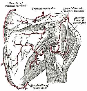
Upper Limb Anatomy
06/11/2009 01:04:46 م
In This Section you will find detailed different Sections about the different organs and structures in the region of the Upper limbs including The shoulder , the arms , the forearms , the hand anatomy... More Details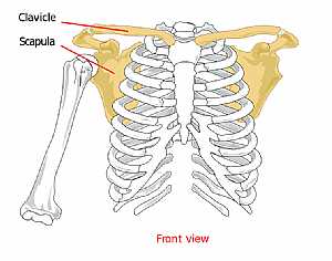
Pectoral anatomy
04/12/2009 06:50:00 ص
this image shows the bones forming the pectoral girdle from anterior view (the clavicle and scapula bone) displaying their position and relation to the rest of the body bones... More Details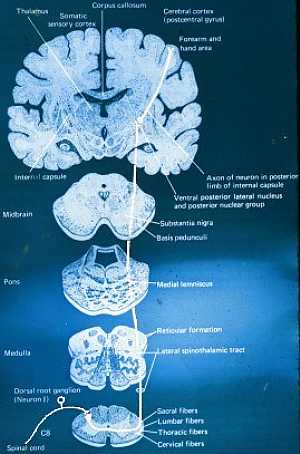
Pathways of C.N.S
22/10/2009 01:52:27 م
In This Section you will find detailed different Photos and images about the anatomy of the Pathways of the CNS including their types , spinothalamic track anatomy , ascending tracks anatomy , descend... More Details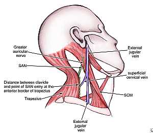
Accessory nerve anatomy
29/10/2009 03:42:00 م
this image shows the spinal part of the accessory nerve passing in the posterior triangle of the neck showing: 1. Great auricular nerve 2. Spinal accessory nerve 3. trapezius m. 4. external jugular n... More Details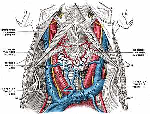
Neck anatomy
11/10/2009 04:02:00 م
this image shows the major vessels in the neck region and their relations to each other showing: 1. sternothyroid m. 2. inf. thyroid vein 3. middle thyroid vein 4. cricothyroid m. 5. sternothyroid art... More Details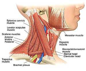
Neck Anatomy
11/10/2009 03:40:00 م
this is the muscles of the neck showing: 1. splenius cervicis m. 2. levator scapulae m. 3. masseter m. 4. digastric m. 5. sternocleidomastoid m. 6. brachial plexus 7. trapezius m. 8. ant. , middle and... More Detailsorthopaedic joint assessment centr dr mcmahon
, , , , , , ,anatomi ligamen panggul wanita
, ,abdomen sans preparation normale
, , , , , ,world conferences on urine therapy
, , , , , , , , , ,© Copyright 2001-2022 eDoctorOnline.com